Pages

Sunday, April 30, 2017

Managing Hypertensive Emergency


A hypertensive emergency (formerly called “malignant hypertension”) is severe hypertension (high blood pressure) with acute impairment of one or more organ systems (especially the central nervous system, cardiovascular system and/or the renal system) that can result in irreversible organ damage. In a hypertensive emergency, the blood pressure should be substantially lowered over a period of minutes to hours with an anti hypertensive agent.

Definition
The term hypertensive emergency is primarily used as a specific term for a hypertensive crisis with a diastolic blood pressure greater than or equal to 120 mmHg and/or systolic blood pressure greater than or equal to 180mmHg. Hypertensive emergency differs from hypertensive crisis in that, in the former, there is evidence of acute organ damage.

Laboratory Evaluation

Obtain electrolyte levels, as well as measurements of blood urea nitrogen (BUN) and creatinine levels to evaluate for renal impairment. A dipstick urinalysis to detect hematuria or proteinuria and microscopic urinalysis to detect red blood cells (RBCs) or RBC casts should also be performed

A complete blood cell (CBC) and peripheral blood smear should be obtained to exclude microangiopathic anemia, and a toxicology screen, pregnancy test, and endocrine testing may be obtained, as needed.

Management

If a patient presents to the emergency department with a high B.P the role of the treating physician is to determine either the patient is exhibiting any signs of end organ damage or not.

Thus, optimal control of hypertensive situations balances the benefits of immediate decreases in BP against the risk of a significant decrease in target organ perfusion. The emergency physician must be capable of appropriately evaluating patients with an elevated BP, correctly classifying the hypertension, determining the aggressiveness and timing of therapeutic interventions, and making appropraite decisions.

Acutely lowering the B.P in some situations may have an adverse effect too.

Pharmacological measures
An important point to remember in the management of the patient with any degree of BP elevation is to “treat the patient and not the number.” In patients presenting with hypertensive emergencies, antihypertensive drug therapy has been shown to be effective in acutely decreasing blood pressure.

Acute pancreatitis


Pancreatitis
is an inflammatory process in which pancreatic enzymes autodigest the gland. Acute pancreatitis is sudden swelling and inflammation of the pancreas. In acute cases the gland heals without any impairment of function or any morphologic changes.

Acute pancreatitis can have severe complications and high mortality despite treatment. While mild cases are often successfully treated with conservative measures.

Etiology
The pancreas is an organ located behind the stomach that produces chemicals called enzymes, as well as the hormones insulin and glucagon. Most of the time, the enzymes are only active after they reach the small intestine, where they are needed to digest food.

When these enzymes somehow become active inside the pancreas, they eat (and digest) the tissue of the pancreas. This causes swelling, bleeding (hemorrhage), and damage to the pancreas and its blood vessels.

Acute pancreatitis affects men more often than women. Certain diseases, surgeries, and habits makes more likely to develop this condition.

The condition is most often caused by alcoholism and alcohol abuse (70% of cases in the United States). Genetics may be a factor in some cases. Sometimes the cause is not known, however.

Other conditions that have been linked to pancreatitis are:
  • Autoimmune problems (when the immune system attacks the body)
  • Blockage of the pancreatic duct or common bile duct, the tubes that drain enzymes from the pancreas
  • Damage to the ducts or pancreas during surgery
  • High blood levels of a fat called triglycerides.
  • Injury to the pancreas from an accident
Other rare causes may include:
  • Complications of cystic fibrosis
  • Hyperparathyroidism.
  • Reye syndrome
  • Use of certain medications (especially estrogens, corticosteroids, thiazide diuretics, and azathioprine)
  • Viral infections, including mumps coxsackie B, and campylobacter.

Examining a Diabetic Patient


Diabetes mellitus is a clinical syndrome characterized by chronic hyperglycemia and disturbances in carbohydrate,lipid and protein metabolism. A patient with diabetes need a complete physical examination to look for any related complications and to manage accordingly.

General Appearance
  • Check the weight and look for obesity and measure BMI
  • Facial appearance: note any features of cushings disease
  • State of hydration
Lower Limbs
  • Inspect and look for any ulceration, infection, hair loss, skin atrophy.
  • Check injection sites on the thigh and note any skin atrophy due to insulin use.
  • Palpate all the peripheral pulses that includes femoral, popliteal, posterior tibial, dorsalis pedis.
  • Check pedal edema that can be present due to nephropathy.
  • Feel the tempreture as the feet may be cold due to ischemia.
  • Do the neurological assessment by checking the sensations including those of dorsal column.
  • Reflexes may be diminished due to neuropathy and also look for any signs of proximal muscle wasting.
  • Joints for Charcot’s joints ( loss of proprioception )
Upper Limbs
  • Nails : for candidiasis
  • Feel upper limb injection sites
  • Check Blood pressure in supine and standing to detect autonomic neuropathy
  • Palpate upper limb pulses .

Mitral Valve Prolapse


Mitral valve bulging back into the left atrium during systole is called mitral valve prolapse. This is also called floppy mitral valve or myxomatous mitral valve.

Etiology
MVP exhibits a strong hereditary component and in some patients is transmitted as an autosomal dominant trait.

Second common cause is rheumatic fever mostly seen in developing countries.

It is twice as frequent in women as in men.

MVP may occasionally result from Marfan syndrome, Ehler danlos syndrome and osteogenesis imperfecta.

Pathology
It develops due to myxomatous degeneration of mitral valve in which middle layer of valve leaflet composed of loose, myxomatous material is unusually prominent.

Clinical Features
Symptoms
  • Most patients with mitral valve prolapse are asymptomatic.
  • In symptomatic patients it may present as chest pain, dyspnea, fatigue, palpitation, syncopeand even sudden death.
  • Spontaneous rupture of chordea tendinae may cause a sudden worsening of MR that is hemodynamically severe.

Introduction to Von Willebrand’s Disease


Von Willebrand’s disease
is the most common congenital disorder of hemostasis. It is transmitted in an autosomal dominant pattern. It is characterized by deficient or defective von willebrand factor. Von willebrand factor plays a role in platelet adhesion to damaged subendothelium as well as stabilizing factor VIII in plasma.

Clinical Manifestations
  • It can affect both males and females.
  • Most bleeding is mucosal ( epistaxis, gingival bleeding, menorrhagia and GI bleeding ).
  • Bleeding follows minor trauma or surgery.
  • Aspirin increases bleeding while pregnancy and estrogen use decreases bleeding.
Investigations
  • Bleeding time : Increased
  • PT : normal
  • APTT : raised
  • Factor VIII : low
  • vWF : low
In von willebrand’s disease bleeding time is prolonged while in Hemophilia bleeding time is normal.

Important Neurological Investigations


There are a large number of neurological disorders and so a number of different investigations are available to help reach the proper diagnosis and then accordingly a proper management.

Here we can discuss a few important ones:

X- Ray Skull
It may show
  • Fracture of skull valut or base.
  • Enlargement or destruction of pituitary fossa due to tumor or raised intracranial pressure.
  • Intracranial calcification due to tuberculoma.
X- Ray Spinal cord
It may show fractures, spondylosis ( degenerative bone disease ) or any destructive bone lesion due to infection or metastasis.

Computed Tomography ( CT Scan )It is an x-ray slice of 5-10 mm thickness in which different tissues have different densities.

Hypodense tissues ( looking blackish )
  • Infarction, tumor, abscess(having white rim and black center), cerebral edema, encephalitis and resolving hematoma.
  • CSF and air looks blacker.

Hyperdense tissues ( looking Whitish )
  • Bone, blood(hemorrhage) and calcifications.
Advantages of CT scan
  • CT scan easily demonstrates
  • Cerebral tumors
  • Intracerebral hemorrhage and Infarction
  • Subdural and Extradural hematoma
  • Subarachnoid hemorrhage
  • Lateral shift of midline structures
  • Cerebral atrophy
  • Pituitary mass lesion

Brief Summary Of Hepatic Encephalopathy


Hepatic encephalopathy
which is also known as portosystemic encephalopathy may be defined as a condition in which there is worsening of normal brain functioning as the liver is not able to detoxify the harmful substance in the blood and is characterized by altered level of consciousness and disorientation.

Pathology
The blood bypasses the liver via the collaterals and the toxic metabolities (e.g ammonia, free fatty acids and mercapts ) pass directly to the brain to produce the encephalopathy. There is also an increased sensitivity of CNS neurons to the inhibitory neurotransmittor (GABA) and an increase in circulating levels of endogenous benzodiazepines. Cerebral edema is frequently present and contributes to the development of clinical features.

Precipitating Factors
  • Increased Nitrogen Load
  • Gastrointestinal bleeding
  • Excess dietray protein
  • Azotemia
  • Constipation
  • Electrolyte and Metabolic Imbalance
  • Hypokalemia
  • Alkalosis
  • Hypoxia
  • Hyponatremia
  • Drugs
  • Narcotics, Tranquilizers. sedatives
  • Diuretics
  • Miscellaneous
  • Infection
  • Surgery
  • Portosystemic shunt
  • Superimposed acute liver failure
  • Progressive liver disease
Clinical Manifestations
Symptoms
  • Disturbance of sleep with sleeping during day and awaking in night is one of the earliest features.
  • Alteration in personality, mood disturbances, confusion, deterioration of self care and deterioration of hand writing, slurring of speech, disorientation, drowsiness and eventually coma develop.
  • Convulsions also sometimes develops.
  • Hyperventilation, fever, nausea and vomiting are also common

Causes Of Dementia


Definition: Dementia may be defined as acquired deterioration in brain’s neuronal function that is characterized by impaired performance of daily living activities including disturbances of memory, thinking, orientation, comprehension, learning and judgement. Memory is the most common cognitive ability lost with dementia.

Causes: Dementia has many causes and it is important to identify and exclude the treatable causes.

A list of different causes of dementia is given in a simplified way as follows:

Most Common Causes To Be Listed First
  • Alzheimer’s disease
  • Vascular Dementia
  • Alcoholism
  • Parkinson’s disease
  • Drug/Medication intoxication
Causes Listed According To Different Etiologies

1. Degenerative
  • Alzheimer’s disease
  • Pick’s disease
  • Huntington’s disease
  • Parkinson’s disease
  • Normal pressure Hydrocephalus
2. Vascular
  • Cerebrovascular disease
  • Cranial arteritis
  • Multi infarct dementia
  • Cardiac arrest

Introduction To Panic Disorder


Panic disorder is an anxiety disorder that is characterized by sudden attacks of fear and panic. A panic attack may be defined has a sudden, intense experience of fear coupled with an overwhelming feeling of danger and death.

Incidence
It occurs in 1-3 % of the population ; female to male ratio is 2 : 1. Familial aggregation is common. Onset is usually in the second or third decade of life.

Clinical Presentation
Initial presentation is almost always to a non psychiatric doctor , frequently in the ER as a possible heart attack or serious respiratory problem. The disorder is often intially unrecognized or misdiagnosed.

Attacks are sudden, unexpected paroxysms of terror and apprehension with multiple associated somatic symptoms.

Usually reach a peak within 10 minutes , then slowly resolve spontaneously.

Panic disorder is often associated with concomitant major depression.

Diagnostic Criteria for Panic disorder The diagnostic criteria for panic disorder requires four or more panic attacks within 4 weeks occurring in non threatening or non exertional settings and attacks must be accompanied by at least 4 of the following ;
  • Dyspnea
  • Palpitations
  • Chest pain or Discomfort
  • Feeling of being choked
  • Dizziness/ vertigo/ unsteady feelings
  • Sweating
  • Faintness
  • Trembling.
  • Fear of dying
  • Going crazy or doing something uncontrolled.

Management Of Patient With GIT Bleeding


A patient with upper or lower GI bleeding after initial evaluation should be managed as follows:

1. Provide venous access with large bore IV (14-18 gauge); central venous line for major bleed and patients with cardiac disease; monitor vital signs, urine output. hematocrit. Gastric lavage is of unproven benefit but clears the stomach before endoscopy. Iced saline may lyse clots ; room temperature tap water may be preferable . Intubation may be required to protect airway.

2. Type and cross match blood (6 units may be needed for a major bleed).

3. Prepare the surgical team for standby when bleeding is massive.

4. Support blood pressure with isotonic fluids (normal saline); albumin and fresh frozen plasma in cirrhotics. Packed red blood cells when available (whole blood if massive bleeding) ; maintain Hct >25-30.

5. IV calcium (e.g upto 10-20 ml 10% calcium gluconate IV over 10 to 15 minutes) if serum calcium falls due to transfusion of citrated blood.

6. Start empirical drug therapy with antacids , H2 receptor blockers, omeprazole although they are of unproven benefit.

A Brief Discussion On Paget’s disease


Paget’s disease
is the disease of the bone caused by a high bone turnover and disorganized osteoid formation. Involved bone is deformed and weak.

How common is paget’s disease
Radiographic evidence is seen in around 2.5% of men over 55 years of age and a little less in women. Less than 10% of those have symptoms. It occurs predominately in people of English origin, including those in North America and Australia. It may be declining in prevalence.

Pathology of bone in paget’s disease.
Paget’s disease is caused by focal or multifocal areas of bone resorption by large osteoclasts, followed by increased bone formation, but the new bone is abnormal with a mosaic appearance on microscopy. It is expanded in size, deforms and fractures more easily and is highly vascularised.

Serum and urine biochemical changes seen in paget’s disease
  • Raised alkaline phosphatase.
  • Raised serum osteocalcin ( not routinely used )
  • Raised urinary hydroxyproline and pyridinolone.
Some other causes of raised alkaline phospahtase

Approach To A Patient With Dysphasia


Dysphasia
is a disorder of language and refers to the inability to understand or find words due to a lesion in the dominant (usually left) hemisphere.

Physiology

Wernicke’s area recognizes sound as a language , but a higher concept area is required to convert sound into meaning. This concept area is then connected to Broca’s area where speech is generated. A direct connection , the arcuate fasciculus also exists between Wernicke’s area and Broca’s area.

Types Of Dysphasia

Receptive Dysphasia: The patient is unable to understand; his speech is fluent but words are meaningless. The lesion is in the Wernicke’s area in the temporal lobe.

Expressive Dysphasia: The patient is unable to express despite comprehension. Can cause frustration to patients who know in their minds what to say but cannot get the words out. Due to lesion in the Broca’s area in the frontal lobe.

Transcortical sensory dysphasia: It is similar to receptive dysphasia but with preserved repetition and is cause by lesion in the parietal-occipital concept area.

Transcortical motor dysphasia: It is similar to expressive dysphasia but with preserved repetition and is caused by incomplete lesion in Broca’s area.

Conductive Dysphasia: Refers to preserved comprehension and output with loss of repetition and is the result of a lesion in the arcuate fasciculus.

Introduction To Ankylosing Spondylitis


Introduction:
This is a chronic inflammatory arthritis especially affecting sacroiliac joints and spine and characterized by progressive stiffening and fusion of the axial skeleton. It is one of the seronegative spondyloarthropathies.

Incidence:  It is most commonly seen in the age range of 20 to 30 years and male to female ratio is   4: 1 . More than 90% of affected persons carry the histocompatibilty antigenHLA B27

Clinical Signs and Symptoms
Onset is usually insidious. There are recurring episodes of low back pain and stiffness sometimes radiating to the buttocks or thigh. Pain is worse in early morning and after inactivity.

Some patients complian of chest pain aggravated by breathing that is due to involvement of costovertebral joints. There is occasionally heel pain due to plantar fasciitis .

On examination there is failure to obliterate lumbar lordosis on forward flexion. There is pain on sacroilliac compression and tenderness over bony prominenece such as illiac crest, ischial tuberosity and greater trochanter.

There is restriction of movement of lumbar spines in all directions. As the disease progresses stiffness increases throughout the spine.

Associated Clinical findings
  • Iritis occurs in about 25% of patients.
  • Aortic regurgitation, heart blocks and anterior uveitis
  • Pulmonary fibrosis of the upper lobes with progression to cavitation mimicking tuberculosis.
Investigations
  • ESR is often raised.
  • RA factor is absent.
  • HLA B 27 seen in 90% of cases
X Ray Lumbar spine

Treatment For Cystic Fibrosis


Cystic fibrosis
is a genetic disorder that is inherited in an autosomal recessive pattern and is characterized by a defective transport of chloride and sodium across the epithelium that causes thick, sticky mucus to build up in the lungs, digestive tract, and other areas of the body. It is one of the most common chronic lung diseases in children and young adults.

Some of the treatment options are discussed briefly here:

1. Physiotherapy
Daily chest percussion and physiotherapy with postural drainage reduces respiratory exacerbations

3. Antibiotics
Early high dose , broad spectrum , long duration (minimum 2-3 weeks) antibiotics helps minimize lung damage in infective exacerbations. Oral or intravenous fluroquinolones combined with nebulized antibiotic is often the initial choice; resistance and chronic carriage needs intravenous combinations. Prophylactic antibiotics are also increasingly used.

3. Bronchodilators
These are helpful for symptomatic relief.

4. DNase- alpha mucolytics
These are administered as areosol spray. Mechanism of action is that it interferes with sputum neutrophil DNA, helping to liquefy sputum and encourage expectoration, thus helps in reducing cough.

Friday, April 28, 2017

Brief Summary of Polycystic Ovarian Syndrome


Polycystic ovary syndrome (PCOS) is one of the most common female endocrine disorders.These patients have a steady state of relatively high estrogen, androgen and LH levels rather than the fluctuating levels seen in ovulating women.

Definition
There are two definitions or diagnostic criteria that are commonly used:

 - In 1990 a consensus workshop sponsored by the NIH(National Institutes of Health)/NICHD(National Institute of Child Health and Human Disease) suggested that a patient has PCOS if she has all of the following:
  • Oligoovulation
  • Signs of androgen excess (clinical or biochemical)
  • Other entities are excluded that can result in menstrual irregularity and hyperandrogenism.
 - In 2003 a consensus workshop sponsored by ESHRE(European Society for Human Reproduction and Embryology)/ASRM(American Society for Reproductive Medicine) in Rotterdam indicated PCOS to be present if any 2 out of 3 criteria are met.
  • Oligoovulation and/or anovualation manifested as oligomenorrhea or amenorrhea
  • Excess androgen activity (clinical or biochemical evidence)
  • polycystic ovaries (as seen on gynecologic ultrasound)
Pathophysiology
Increased level of estrogen comes from obesity due to conversion of ovarian and adrenal androgen to estrone in body fat. High estrogen level suppresses FSH and causes relative increase in LH. Constant LH stimulation of ovary results in anovulation, multiple cysts and theca cell hyperplasia with excess androgen production.

Conditions Causing Skin Hyper pigmentation


Sometimes in medical practice patients presents with skin hyperpigmentation or pigmented spots. There are many underlying different causes and they are briefly discussed here:

1. Endocrine Disorders
  • Addison’s disease
  • Cushing’s syndrome
  • Acromegaly
  • Nelson’s syndrome
  • Pregnancy
  • Porphyria
2. Renal Failure

3. Cirrhosis

4. Haemochromatosis

5. Nutritional Causes:
  • Vitamin B12 deficiency
  • Pellagra
6.. Amyloid

7. Acanthosis nigricans

Thursday, April 27, 2017

Transient Ischemic Attacks


Definition:
Transient Ischemic Attack is defined as a condition in which the symptoms of stroke are lasting less than 24 hours. The symptoms usually reach their peak in seconds and lasts for minutes or hours (but by definition less than 24 hours).

TIA has a tendency to recur and to predispose to a complete stroke. There is a complete functional recovery after a TIA and the focal neurological deficits during the episode depend on the artery involved.

Etiology: Transient ischemic attacks are caused by the passage of emboli in about 90% of cases and less commonly it is due to a fall in cerebral perfusion (e.g due to a cardiac dysrhythmia, postural hypotension or reduced blood flow through atheromatous vertebral arteries).

The principal source of emboli is atheromatous plaque within the carotid or vertebral arteries or from mural thrombus formed on diseased heart muscle.

Clinical features
  • TIA’s cause sudden loss of function in one region of the brain depending on the artery involved.
  • Consciousness is usually preserved.
  • On examination of the patient there may be flaccid weakness, sensory loss, hyperreflexia and extensor plantar response on the affected side.
  • Sources of emboli may be evident such as valvular heart disease, endocarditis, recent MI or atrial fibrillation.
  • Associated diseases may be diagnosed like atheroma, hypertension, braydycardia or diabetes.
  • Sometimes patients present with transient monoocular blindness termed as Amaurosis fugas.
  • Some patients may experience episode of amnesia with confusion lasting for several hours caused by ischemia of posterior circulation.

A Brief Discussion On Urinalysis


Urinalysis means examining or testing the urine and it is one of the most common and easy method used for medical diagnosis. It involves the physical, chemical, and microscopic examination of urine.

A part of a urinalysis can be performed by using urine test strips, known as Dipstick in which the test results can be read as color changes. Another method is examination of the urine sample under light microscopy.

Physical Or Macroscopic Examination Of urine
The first part of a urinalysis is direct visual observation:

1. Colour
Normal color of the urine ranges from colorless to deep yellow depending on the concentration of the urochorme pigment. Different colors may be observed in different conditions like red with blood in the urine or certain food colors e.d beetroot. orange with rifampicin, yellow in jaundice or dehydration and black with severe hemoglobinuria.

2. Volume
Normal volume is 800-2600 ml/day and it is referred to be oligouria if its < 300 ml/day and anuria if < 100 ml/day.

3. Appearance
A normal urine sample appears clear on visual inspection. Turbidity or cloudiness may be caused by excessive cellular material or protein in the urine or may develop from crystallization or precipitation of salts upon standing at room temperature or in the refrigerator. Clearing of the specimen after addition of a small amount of acid indicates that precipitation of salts is the probable cause of tubidity.

Introduction to Renal Biopsy


Renal biopsy is not a commonly performed procedure in medical practice but it is sometimes indicated when it has diagnostic value in certain kidney diseases. Most cases of acute renal failure are due to pre renal causes or acute tubular necrosis and recovery of renal function typically occurs over a course of few weeks. Renal biopsy should only be performed if knowing the histology will help in the management. Once chronic renal failure is established, the kidneys are small, may be hard to biopsy, and theresults are usually unhelpful.

Indications
  • To determine the cause of unexplained renal failure.
  • Persistent Heavy proteinuria >1gm/24hrs
  • Investigating the cause of adult nephritic syndrome with persistent hematuria.
  • Childhood nephritic or nephrotic syndrome that are unresponsive to treatment.
  • Systemic disease with renal involvement like sarcoidosis, amyloidosis.
  • Post transplant renal dysfunction.
Contraindication
  • Underlying disorder of coagulation.
  • Thrombocytopenia.
  • Uncontrolled hypertension.
  • Solitary kidney (except in transplanted kidneys).
  • Small contracted kidneys i.e < 60% of expected bipolar length because it is technically difficult, histology is hard to interpret and prognosis cannot be altered.
  • Uncooperative patient.

Introduction To Analgesic Nephropathy


Introduction: 
Analgesic Nephropathy is a condition that is associated with prolonged, heavy use of analgesics that leads to decline in renal function due to chronic papillary necrosis and chronic diffuse tubulointerstitial damage to renal cortex.

The incidence of analgesic nephropathy has declined significantly after the withdrawal of phenacetin use.

Clinical Features
  • Patients with analgesic nephropathy are unable to produce maximally concentrated urine due to underlying medullary and papillary damage.
  • They present with repeated UTI’s and or urinary abnormalities on urinalysis like hematuria, proteinuria, casts etc.
  • Anemia is more severe as compared to level of renal insufficiency.
  • Distal tubular acidosis associated with analgesic nephropathy contributes to the development of nephrocalcinosis.
  • Kidneys become shrunken and calyces are deformed producing “ring sign” on IVP that is pathognomonic of papillary necrosis.
  • Transitional cell carcinoma may develop in the urinary pelvis or ureters as a late complication of analgesic abuse.

Wednesday, April 26, 2017

Brief Summary About Anal Fissure

Definition: An anal fissure is a cutaneous tear in the ectodermal portion of the anal canal and is usually found in the postero-lateral wall or in other words at 12 o’clock or 6 o’clock positions around the anal circumference.

Clinical Features
  • It commonly occurs after a bout of constipation and is very painful.
  • Patients find themselves in a vicious circle where they appreciate that the next bout of defecation will be painful, hence they avoid passing a stool and progressively become more constipated.
  • When the bowel is eventually opened the tear is made more worse.
  • Fresh bleeding is common and this can be a cause of bleeding per rectum in many patients.
Clinical Diagnostic Feature
A diagnostic feature of an anal fissure is anal pain after defecation and in chronic cases the skin at the lower part of the fissure becomes swollen and can be used as a marker of an anal fissure- the sentinel pile. This may be the only sign of a chronic anal fissure and it is often too painful to examine the patient proctoscopically.

Etiology
Anal fissures are very common in young infants, but may occur at any age. The rate of anal fissures drops with age. Fissures are much less common among school-aged children than infants.

In adults, fissures may be caused by passing large, hard stools, or by having diarrhea for a long time. 

Other factors may include:
  • Decreased blood flow to the area in older adults
  • Too much tension in the sphincter muscles that control the anus

Treatment Options For Rheumatoid Arthritis

Patients with rheumatoid arthritis need proper management and control of their symptoms to improve the quality of life. Although taking analgesic medicines for a long duration of time leads to gastric problems but it is important to give patient some relief from disturbing pain and inflammation.


Non Pharmacological Management
  • Encourage regular exercise, physio and occupational therapy.
  • Provide household aids and personal aids e.g wrist splints.
  • Good mental and social support to improve life style and cope up with the disease.
Pharmacological Management
NSAIDs
If there is no contraindication e.g an active peptic ulcer start with an NSAID, such as ibuprofen. There is no single NSAID that is superior to other agents, and newer agents have not been shown to have a decreased incidence in toxicity. If patient cannot tolerate NSAIDs then consider COX2 inhibitors that prevents gastrointestinal side effects.

Steroids
Steroids may decrease joint damage and control difficult symptoms but using them for prolonged period of time leads to decreased bone density and predisposition to osteoporosis as well as other side effects related to prolonged steroid use.

Approach to Patient With Amenorrhoea


It is common in general practice that patients may come with amenorrhoea and it needs a proper diagnostic approach.

Primary Amenorrhoea
Defined as when a girl have never started menstruation and sexual characteristics have not developed by the age of 14 years OR no menstruation by age 16 years when growth and sexual development is normal.

Causes
1. Outflow Abnormalities: Mullerian agenesis, transverse vaginal septum, androgen insensitivity, imperforate hymen.

2. Ovarian Disorders: Gonadal dysgenesis due to chromosomal abnormalities e.g Turner’s syndrome

3. Pitutary Disorders: Prolactinoma

4. Hypothalamic disorders: Kallman’s syndrome.

Secondary Amenorrhoea
Defined as absence of menses for more than 6 months in a previously menstruating woman.

Causes

1. Always consider the possibility of pregnancy.

2. Stress

3. Starvation , anorexia or excessive exercise.

4. Hypo/ Hyperthyroidism

5. Hyperprolactinaemia

Diabetes- Patient Monitoring,Care And Education


Diabetes mellitus is a common disease affecting a large number of population. It is characterized by increased blood sugars and abnormalities of carbohydrate and lipid metabolism.Diabetes can be managed by family physicians as effectively as in hospital clinics.

Aims Of Diabetic Care
  • Alleviation of symptoms.
  • Minimization of complications.
  • Reduction of early mortality.
  • Quality of life enhancement.
  • Education of the patient and family.
  • Features of well organized care
  • Use of a register and well maintained records.
  • Regular review , following a proper protocol and provision of adequate time and open access for patients to review advice.
  • Multidisciplinary team covering all aspects of diabetes care- GPs, diabetes nurses/assistants and educators.
  • Continuing education for professional staff.
Routine diabetic review
Each diabetic patient requires 6 monthly review or more frequently as necessary. This should include a through annual review of all aspects of disease and care.

Review include

Causes Of Dysphagia


Dysphagia means difficulty in swallowing. It may or may not be associated with pain on swallowing. Dysphagia may be associated with ingestion of solids or liquids or both. It is important to know different medical conditions that lead to dysphagia and a simple list is given here:

Causes Of Dysphagia

Congenital
  • Oseophageal atresia
Acquired

1.In the lumen
  • Food bolus
  • Foreign body
2. In the wall
  • Inflammatory stricture
  • Gastroesopahgeal reflux
  • Caustic stricture
  • Candidiasis
  • Achlasia
  • Carcinoma
  • Plummer vinson syndrome
  • Irradiation
  • Scleroderma
  • Chagas disease (rare)

Approach To Elderly Patient


Family physicians are responsible for caring increasing number of elderly patients.Older patients have co morbidities, functional, psychological and social problems that need to be considered by the the family physician.

Goals of Geriatric Assessment

1. Focus on preventive medicine rather than acute medicine.
2. Focus on improving or maintaining functional ability and not necessarily on a “cure.”
3. Provide a long-term solution for “difficult to manage” patients with multiple physicians, recurrent emergency department visits, and hospital admissions with poor follow-up.
4. Aid in the diagnosis of health-related problems.
5. Develop plans for treatment and follow-up care.
6. Establish plans for coordination of care.
7. Determine the need and site of long-term care as appropriate.
8. Determine optimal use of health care resources.
9. Prevent readmission into the hospital.

The geriatric assessment can be divided into four categories: medical, functional, psychological, and social.

Medical Assessment The medical assessment includes a review of the patient’s medical record, medication history (past and present), and a nutritional evaluation. On average, elderly patients have four to six diagnosable disorders, which may require the use of several medications. One disorder can affect another, and in turn a collective deterioration of both can lead to overall poor outcomes.

During the medical assessment, the review of systems should be completed with special emphasis on sensory impairment, dentition, mood, memory, urinary symptoms, falls, nutrition, and pain.

A Brief Summary Of Insomnia


Definition

Insomnia is defined as inadequate or poor-quality sleep characterized by one or more of the following: difficulty falling asleep, difficulty maintaining sleep, waking up too early in the morning, or sleep that is not refreshing. Insomnia also involves daytime consequences such as fatigue, difficulty concentrating, and irritability.

Acute Insomnia : Periods of insomnia lasting between 1 night and a few weeks are defined as acute insomnia.

Chronic Insomnia : Chronic insomnia refers to sleep difficulty occurring at least 3 nights per week for 1 month or more.

Causes
Insomnia may be associated with specific sleep disorders, including restless legs syndrome, periodic limb movement disorder, sleep apnea, and circadian rhythm sleep disorders.

Restless legs syndrome is characterized by unpleasant sensations in the legs or feet that are temporarily relieved by movement. Symptoms are worse in the evening, especially when a person is lying down and remaining still. The sensations cause difficulty falling asleep and are often accompanied by periodic limb movements.
Periodic limb movement disorder is characterized by bilateral repeated and rhythmic, small-amplitude jerking or twitching movements in the lower extremities and, less frequently, in the arms. These movements occur every 20 to 90 seconds and can lead to awakenings, which are usually not noticed by the patient. Often the patient reports that sleep is not refreshing. In many cases, the bed partner is more likely to report the movement problem.

Introduction To SLE


Definition

Systemic lupus erythematosus (SLE) is a chronic inflammatory autoimmune disorder that affects the connective tissues. SLE is characterized by recurring remissions and exacerbations, which are especially common during the spring and summer.

Incidence
It strikes women 8 times as often as it does men, increasing to 15 times as often during childbearing years.

Etiology: 
The direct cause is unknown but it can be predisposed by certain factors
Predisposing factors
  • Physical or mental stress
  • Streptococcal or viral infections
  • Exposure to sunlight or ultraviolet light
  • Immunization
  • Pregnancy
  • Abnormal estrogen metabolism
  • Drugs, including procainamide, hydralazine, anticonvulsants; less commonly, penicillins, sulfa drugs, oral contraceptives
Pathophysiology
Autoimmunity is believed to be the prime mechanism in SLE. The body produces antibodies against components of its own cells, such as the antinuclear antibody (ANA), and immune complex disease follows. Patients with SLE may produce antibodies against many different tissue components, such as red blood cells (RBCs), neutrophils, platelets, lymphocytes, or almost any organ or tissue in the body.

Differential Diagnosis Of Joint Disorders


Disorders of joint is one of the common problem a family physician comes across. Here is a list of differential diagnosis for joint disorders with brief description of each disorder:

1. Congenital Causes

Achondroplasia: Premature osteoarthritis may occur in achondroplasia.

Ehlers-Danlos syndrome: a genetic disorder of connective tissues in which patients have hypermobile joints and hyperextensible skin. they may present with joint subluxation, dislocations and swelling due to effusions.

Marfan Syndrome: Patients have generalized joint laxityand present with joint pain and swelling due to effusions.

Osteogenesis Imperfecta: Patients have brittle bones that lead to easy joint dislocations.

2. Accquired Causes

Infective:
  • Septic or pyogenic arthritis usually present as a monoarthritis with a red , swollen, painful, immobile joint. Usually a single joint is involved.
  • Viral arthritis may occur following viral illness like rubella, mumps, hepatitis etc.
  • In rheumatic fever which usually follows a streptococcal infection. There is a migratory poly arthritis together with carditis, erythematous skin lesions and sub cutaneous nodules.
 Inflammatory:
  •  Rheumatoid arthritis presents initially with swollen, painful, stiff hands and feet. Later characteristic deformities develop. Most commonly swelling is seen at metacarpophalangeal joints.
  • Psoriatic arthropathy usually follows several years after skin lesions.
  • Reiter’s disease may present with arthritis in a single or few joints. There will be a history of sexual contact, urethritis and conjunctivitis.
  • Ankylosing spondylitis presents in young males and initially presents with morning stiffness in the spine. The sacroilliac joints are involved.
  • SLE presents with malaise arthralgia, myalgia, a butterfly rash on the face, raynaud’s syndrome, splenomegaly, pleurisy, pleural effusion, pericarditis.
  • Polymyalgia rheumatica is associated with tender muscles, mild polyarthritis and there may be associated temporal arteritis.

Approach To A Patient Presenting With Hoarseness


Definition Of Hoarseness

Hoarseness is a change in the pitch or quality of the voice caused by abnormalities of vocal cords.

Etiology

Local Causes:
  • URTI ( most common cause )
  • Laryngitis
  • Trauma to the vocal cords: shouting, coughing, vomiting, instrumentation.
  • Hypothyroidism
  • Acromegaly
  • Chronic acid reflux
  • Allergies
  • Heavy smoking or drinking or both
  • Carcinoma
Neurological Causes:
  • Laryngeal nerve palsy
  • Motor neuron disease
  • Myasthenia gravis
  • Multiple sclerosis
Muscular Causes:
  • Muscular dystrophy
Functional Cause.

  • Sometimes no definitive organic cause can be identified and it might be due to psychological causes.

Clinical Features Of Patients Presenting With Angina


Definition: 
Angina is defined as a condition in which patient suffers from chest pain due to decreased blood supply to the cardiac muscles. Most common cause is obstruction or spasm of coronary arteries.

Clinical Presentation
Diagnosis is usually made on clinical history. Patients with stable angina present with:

Pain: Episodic central-crushing or band like chest pain that may radiate to jaw/ neck or one/both arms. Pain in the arm/ neck may be the only symptom. It is important to ask about the frequency, severity, duration and timings of the pain.

Precipitating/ Relieving Factors: Angina pain is usually precipitated by exertion, cold, emotion, and/or heavy meals. Pain stops with rest or using glyceral trinitrate.

Associated Symptoms:  May be associated with palpitations, sweating, nausea and /or breathlessness during attacks.

Presence of risk factors: Patients who suffer from angina have certain risk factors like smoking, family history of cardiac disease, history of other vascular disease.

Tuesday, April 25, 2017

Approach To Patients Presenting With Headache


Headache is one of the common presenting complaints in a physicians office. It is important to identify which headaches are benign, needing no intervention and which requires action. Following questions needs to be asked in history:

Clinical History

1. Does the patient have more than one type of headache?

2. Time:When did the headache start? New or recently changed headache calls for especially careful assessment. How often do they happen? Do they have any patteren( e.g constant,episodic,daily). How long do they last? Why is the patient coming to the doctor now?

3. Character: Nature and quality, site and spread of the pain. Associated symptoms e.g nausea/vomiting, visual disturbance, photophobia, neurological symptoms.

4. Cause: Ask about predisposing and /or trigger factors; aggrravating snd /or relieving factors; family history of similar headaches.

5. Response:Details of medication used (type, dose, frequency, timing). What does the pateint do? e.g can the patient continue work?

6. Health between attacks: Do the headaches go completely or does the patient feel unwell between attacks?

7. Anxieties and concerns of the patient

Physical Examination

Investigations Required In Patients With Ascites


Ascites
is defined as collection of excess free fluid in the peritoneal cavity and it could be due to a number of different medical conditions. A proper history and clinical examination may reveal the underlying cause but still certain general and specific investigation are to be carried out in order to confirm or find out the possible etiology.

General Investigations

1. Urine Dipstick: It is a simple and cheap procedure that will be strongly positive for protein in nephrotic syndrome. If so, a 24 hour urine collection for protein should be undertaken; more than 3.5gm is indicative of nephrotic syndrome.

2. Complete Blood Count: Raised white cell count may indicate an infective etiology but a differential white count is more specific.

3. Urea and Electrolytes: Elevated urea and creatinine may indicate a renal etiology; however , it may also be a component of hepatorenal syndrome, which is renal impairment secondary to liver failure.

4. LFT’s : May be deranged in the presence of liver disease. The serum albumin will be able to indicate hypoalbuminaemia but the underlying cause must still be sought.

5. Chest X-ray: Findings suggestive of cardiac failure are cardiomegaly, upper venous diversion of blood, the presence of Kerley B lines, pulmonary edema and pleural effusion. Occasionally the presence of carcinoma may be suggested by a mass in the lung.

6. Ultrasound Abdomen: In addition to confirming the presence of ascites ultrasound will detect any intra abdominal masses that are not palpable on clinical examination. It may also indicate the presence of fatty deposits in the liver in the presence of cirrhosis. Dilated collateral veins may be visualized in conditions that cause obstruction of the venous outflow of the liver, including cirrhosis and Budd-Chiari syndrome.

7. Abdominal Paracentesis: Aspiration of the ascitic fluid is very useful to help determine the underlying cause. A sample should be sent to microbiology clinical chemistry and pathology.

Ascitic fluid Examinations

Thiamine (Vitamin B1) Deficiency and Wernicke-Korsakoff Syndrome


Introduction:
Thiamine is important for the metabolism of carbohydrates, especially glucose, and amino acids. It is a water-soluble vitamin found in meat, especially liver, and unrefined cereals and grains.

Thiamine is absorbed readily in the upper small bowel. Increased requirements are seen in pregnancy, lactation, thyrotoxicosis, and fever. Increased losses are seen with diuretic therapy and peritoneal dialysis.

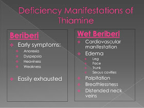
Clinical Features: Thiamine deficiency causes syndromes affecting the cardiovascular and neurologic systems. They are better known by the names beriberi and Wernicke-Korsakoff syndrome. There are two forms of beriberi, wet and dry.

In developed countries, thiamine deficiency is seen mostly in chronic alcoholics. However, it has been reported to occur in others, such as pregnant women with hyperemesis gravidarum on prolonged intravenous fluid therapy, patients on parenteral nutrition, and elderly patients. In alcoholics, thiamine deficiency is due to decreased intake consequent upon a poor diet and also decreased absorption from the intestine.

Neurologic and cardiovascular manifestations are the common presenting symptoms of thiamine deficiency.

Introduction To Vitamin D Deficiency


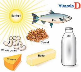
Vitamin D also commonly known as Sunshine vitamin is a fat soluble vitamin and essential to normal body growth and development. It is produced in the body in response to skin being exposed to sunlight. The deficiency of vitamin d may occur due to either inadequate nutritional intake or inadequate sunlight exposure.

Sources Of Vitamin D:There are two forms of Vitamin D known as D2 and D3.
  • Vitamin D2 also known as Ergocalciferol basically comes from fortified foods, plant foods and supplements.
  • VitaminD3 also known as Cholecalciferol comes from fortified foods, Animal sources like fish, eggs and liver as well as can be synthesized in the body when the skin is exposed to ultraviolet rays from the sun.
Symptoms Of Vitamin D Deficiency:
Mild deficiency of Vitamin D is usually not associated with any symptoms. The main symptoms are bone pain and muscle weakness. The deficiency of Vitamin D is associated with a number of different health disorders like:
  • Rickets in children characterized by impaired growth and deformity of long bones.
  • Osteomalacia in adults characterized by muscle weakness and bone fragility
  • Osteoporosis a condition with reduced bone mineral density and increased risk of fractures.
  • Increased risk of death from cardiovascular disease.
  • Cognitive impairment in older adults
  • Muscle twitching or fasciculations .
  • Severe asthma in children
  • Increased risk of cancer.
  • Increased chanced of being diagnosed with Schizophrenia

Managing Vitamin D Deficiency


Deficiency of Vitamin D can affect people of all ages and is usually manifested as bone and muscle pain and weakness. The goal of treatment is to bring the levels to normal, prevent bone fractures and relieve the symptoms. Recent research and evidence shows that vitamin D deficiency is very common in general population. Average daily Vitamin D intake in the diet is usually inadequate to maintain normal blood levels.

Diagnosis Of Vitamin D Deficiency:
A low level of Vitamin D can be diagnosed with a blood test that checks the levels of 25 hydroxyvitamin D:
  • A normal level is defined as a concentration greater than 30 ng/ml
  • Insufficiency is defined as levels b/w 20-30 ng/ml
  • Deficiency is defined as levels less than 20 ng/ml
Management:
The amount of vitamin D that is needed to correct the deficiency depends upon the severity of the deficiency.

Rule of thumb: for every 1 ng/ml increase in your blood level you need an additional 100 IU vitamin D per day.

Prophylactic Antibiotics In Surgery


Whenever surgeries are planned antibiotics are usually given to prevent the risk of wound infections which occurs in appx. 20% of GI surgeries. Prophylaxis is recommended for almost all clean or contaminated and dirty procedures.

Factors supporting the prophylaxis:There are certain patient related or procedure related factors that support the giving of antibiotics in order to prevent infection associated with the surgical procedure. These factors may include:
  • Patients who have some valvular heart problem
  • Immunocompromised patients.
  • Too young or very old age.
  • Procedures involving mouth, GI tract, GU tract and respiratory tract.
Timings Of antibiotic Administration:
It is important to give the antibiotic at a n appropriate time before the surgery in order to give the maximum benefit. The first dose is usually given before the surgery about 30 minutes before the incision. Re administration at one to two half live of the antibiotic is usually recommended.

Antibiotic Regimens: It depends upon personal and local preference but the usual recommendation is as follows:

Monday, April 24, 2017

Depression In Elderly


Depression in elderly is very common but that does not mean that it is normal. It affects about 6 million Americans who are above the age of 65 years. Older patients are more at risk of suffering from depression due to a number of factors like retirement, isolation, death of loved ones and medical problems.

Depression- Definition: Anyone can feel sad or down but when these feelings of sadness are persistent causing trouble in daily routine activities then it becomes a disease. Depression is defined as a medical illness in which there is persistent feeling of sadness, often associated with discouragement and lack of self worth.

Clinical Features:Identifying an elderly suffering from depression is necessary as it is frequently confused with the symptoms of other medical conditions the patient is already having and with the affect of multiple treatments the patient is receiving. It is important to remember that depression is a disease that affects both the body and the mind. The main signs and symptoms are as follows:
  • Sadness
  • Fatigue
  • Lost of interest in hobbies and things that used to give pleasure in the past
  • Decreased appetite
  • Sleep disturbances ( both either difficulty sleeping or excessive sleep )
  • Weight loss
  • Subtle signs of distress like irritable, agitated or difficulty sitting still.
  • Inability to concentrate.
  • Generalized body aches, headaches, digestive disturbance
  • Thoughts of suicide or a suicidal attempt.

Depression In elderly Is Different from Depression In Younger Patients:
  • Depression affects older people differently than in younger patients. Depression in the elderly often increases the risk of cardiac disease by two fold and thus increases the risk of death.

What Is Chemical Pregnancy ?


Introduction:
A chemical pregnancy is actually a very early miscarriage which takes place at a time when ultrasound scan could not show a gestational sac. The only evidence that indicates that the woman got pregnant is that she gets a positive pregnancy test which was done at a very early time.

The excitement of getting a positive pregnancy test usually ends up with getting a period shortly after or b y a negative test when the test is repeated.

Pathophysiology:
A chemical pregnancy occurs when an egg is fertilized but it does not implant on the uterine wall. As a result of fertilization the pregnancy hormone beta HCG starts to be produced in the body. Measuring this hormone in the blood or urine is the first documentation of being pregnant and since a number of pregnancy tests available in the market today can detect this hormone at a very early stage the woman thinks she is pregnant but since the implantation fails this very early miscarriage occurs at a time just around the expected time of periods and without the gestational sac being formed.

Causes:
There are a number of possible cause and is very common and usually goes unidentified since not all woman take a pregnancy test that early.
  • Inadequate uterine lining.
  • Low hormone levels
  • Luteal phase defect
  • Infections
  • Chromosomal abnormalities in the fertilized ovum
  • Unknown reasons

An Approach To Patient With Bleeding


When ever a patient presents with the main complaint of bleeding there are three set of questions that need to be answered ;

1. Is There an Emergency?
A condition that may need and immediate resuscitation or help by a senior. The patient may seem to faint or go into shock. check the blood pressure for hypotension. Consider any signs of CNS bleeding like meningism as it is also an emergency. Also consider an underlying condition in which an apparently minor bleed may evolve into a catastrophic event like bleeding in pregnancy or the puerperium.

2. Why Is the Patient Bleeding?
It is important for a physician taking care of a patient with bleeding to think that is the bleeding with which the patient has presented is normal given the circumstances for example surgery, trauma, postpartum etc or that there is some other reason like bleeding disorder, drugs, liver disease or sepsis that is leading to the problem.

3. What is the mechanism of bleeding If it is a case of bleeding disorder?
To help find the answer some diagnostic tests need to be done like full blood count, coagulation studies, D-dimers, bleeding time etc.

Management:

Grief – How To Deal It In Family Medicine Practice


Introduction

Loosing someone or something that a person really loves and cares for is very heart breaking, painful and emotionally a difficult process to go through. Grief can be defined as an emotional reaction to a significant loss. The term grief and bereavement is usually used in medical practice for a person who has suffered the the death of a beloved one but grief can also be experienced in conditions like when a person is diagnosed with some incurable disease, breakup of a close relationship etc. Grief reaction is normal in all theses circumstances and every individual has its own ways to cope with it and go through it .

Clinical features Of a Grief Reaction:Everyone feels grief in its own way but there are common stages that usually help to describe it. These stages may not occur in a specific order and not everyone experiences all of these emotions.

5 stages of Grief Reaction:

Denial, disbelief and numbness: The first reaction is usually to deny the reality of the situation. It is actually a defense mechanism of the body to protect from the harmful effects of the immediate shock. This emotion is short lived and temporary.

Anger and Blaming others: When the masking effect of denial starts to wear off and the reality begins to get clear the person is still not ready to accept it and it comes out as an emotion of anger. The anger may be aimed at anything, on inanimate objects, friends, family or even strangers. Even the doctor who diagnosed the disease or pronounced the death of the loved one becomes the target and the one to be blamed.

Bargaining: This is a reaction of feeling of helplessness and trying to reverse the situation back to the past. The thought like ‘If we have gained medical attention sooner....’, ‘if we would have taken a second opinion..…’ etc comes in the mind. Secretly the person going through this stage of grief tries to make a deal with God or his higher power that he would do anything just to prevent or reverse the inevitable situation he is grieving for.

Managing High Blood Pressure


Introduction:

Millions of people around the globe have been diagnosed with high blood pressure or hypertension and it is one of the most common risk factor for stroke and heart attack. High blood pressure can also lead to damage to the blood vessels leading to kidney problems, aneurysms as well as damage to the eyes. It is very important to prevent, control and treat hypertension appropriately.

Guidelines For Target Blood Pressure:

The new guidelines recommend the following target for controlling high blood pressure:
  • among adults age 60 and older with high blood pressure, aim for a target blood pressure under 150/90.
  • among adults age 30 to 59 with high blood pressure, aim for a target blood pressure under 140/90
  • among adults with diabetes or chronic kidney disease, aim for a target blood pressure under 130/80.
Management:Managing high blood pressure involves diet changes, exercise, life style changes and if requires certain medications.

1. Loosing extra weight: Blood pressure often increases as weight increases. Loosing just 10 pounds helps reduces the blood pressure. Beside just loosing weight it is important to keep an eye on the waistline since carrying too much weight around the waistline leads to an increase in blood pressure.

2. Regular Exercise: Exercising regularly has a great impact on preventing and controlling high blood pressure. Even moderate activity for 10 minutes at a time or just walking may alos help a lot.

3. Eating A healthy diet: Aim to eat a diet rich in fruits, vegetables, whole grains and high fiber food. avoid fatty foods and also limit sugar intake.

4. Reduce Sodium In the diet: Even a small reduction of sodium in the diet can help reduce blood pressure greatly. Avoid eating processed food like chips and fast food items that are high in sodium content.

Angina And Its Treatment


Angina is a pain that comes from the heart usually as a result of narrowing of the coronary arteries and as a result the heart muscle does not get as much blood and oxygen supply as needed. The pain of angina is described as an uncomfortable pressure, discomfort and squeezing in the center of the chest. Sometimes the pain may be felt in the arm, neck, jaw or shoulder. A number of medications and treatment options are available for managing angina.

Non-Pharmacological Treatment:
  • If the patient smokes it is important to make every possible effort to stop him smoking.
  • The blood pressure should be checked regularly and if high should be manged accordingly.
  • If the patient is over weight , loosing weight is advised.
  • Avoid inactivity and the aim should be a moderate physical activity on most of the days of the week for at least 30 minutes daily.
  • If blood cholesterol is high it should be treated
  • The patient should eat a healthy diet and avoid fatty foods.
  • Avoid drinking alcohol.
  • Avoid taking large heavy meals that may trigger an attack of angina.
  • Patients are advised to slow down or take rest if physical activity triggers an attack of angina.
  • Some patients may get an angina due to a emotional stress so try to avoid the situations that causes stress.
Pharmacological Treatment:

1. Nitrates: are the most common drugs used for the angina treatment. Among the nitrates Nitroglycerin is the most commonly used medicine. Nitrates act by relaxing and dilating the blood vessels. This allows more blood to flow to the heart and also decreases the work load of the heart. Nitroglycerin or glyceryl trinitrate medicine comes in the form of a tablet or a spray and the dose is given under the tongue when the pain of angina develops. The medicine is absorbed quickly into the blood stream and acts within minutes to relieve the pain. The patients with a history of angina or those who are at risk are advised to carry the medicine with them all the time and use it as soon as needed. The most common side effect with this drug is a headache which usually improves with continued use.

Introduction To Sleep Disorders


Introduction:

Sleep disorders may be defined as any condition that prevents a person from getting a restful sleep. Good sleep is necessary for optimal health and normal functioning of the human body. Some sleep disorders may be serious enough to impair the physical and mental functioning of the person.

Normal Sleep:Before different sleep disorders are summarized here it is important to know what is the normal sleep pattern. The amount of sleep a person requires depends on many factors like age, gender, pregnancy etc
  • Infants require 14-15 hrs of sleep a day
  • Teenagers need an average of 8-9 hrs of sleep daily.
  • Most adults need 7- 9 hrs sleep at night but some may need as few as 6 hrs or some may be satisfied after 10 hrs of sleep.
  • Women in the first 3 months of pregnancy need some extra hours of sleep than usual.
Sleep Disorders: A few of the sleep disorders are summarized here :

Insomnia: a hard time falling or staying asleep.

Sleep apnea: a condition in which breathing difficulty interrupts sleep.
Delayed sleep phase disorder: the patient feels really difficult to wake up in the morning and the biological clock is disturbed. It becomes impossible to wake up even with a loud alarm clock or a person waking the patient up.

Brief Summary Of Benzodiazepines


Introduction:

Benzodiazepines are a class of drugs also know as tranquilizers that acts on the central nervous system and are mainly used to treat different anxiety disorders.

Mechanism of action:
Although the exact mechanism of action is not known these drugs effect the GABA neurotransmitter in the brain. The response to this inhibitory neurotransmitter is enhanced and thus it becomes resistant to excitation.

Common Drugs In this Group:
All benzodiazepines are similar in pharmacological action but have different potencies, and some bezodiazepines work better in the treatment of certain conditions than the other. They are used mainly as sedatives, hypnotics, muscle relaxants, anxiolytics and anticonvulsants. Common drugs in this group include:
  • Alpralzolam
  • Lorazepam
  • Midazolam
  • Bromazepam
  • Diazepam etc

Medical Uses: Benzodiazepines may be prescribed for one of these conditions:
  • Anxiety
  • Insomnia
  • Alcohol Withdrawal
  • Seizures
  • Depression
  • Sedation prior to surgery or any diagnostic procedure
  • Panic disorders
  • Muscle spasms
Side Effects: The common side effects may include:

Raynaud’s Disease - Definition, Causes, Symptoms, Diagnosis And Treatment


Introduction
: One of several primary arteriospastic disorders, Raynaud’s disease is characterized by episodic vasospasm in the small peripheral arteries and arterioles, precipitated by exposure to cold or stress. This condition occurs bilaterally and usually affects the hands or, less often, the feet.
Raynaud’s disease is most prevalent in women, particularly between puberty and age 40. A benign condition, it requires no specific treatment and has no serious aftereffects.

Raynaud’s phenomenon, however, a condition often associated with several connective tissue disorders—such as scleroderma, systemic lupus erythematosus, and polymyositis—has a progressive course, leading to ischemia, gangrene, and amputation. 

Differentiating the two disorders is difficult because some patients who experience mild symptoms of Raynaud’s disease for several years may later develop overt connective tissue disease—most commonly, scleroderma.
Causes
Although the cause is unknown, several theories account for the reduced digital blood flow: intrinsic vascular wall hyperactivity to cold, increased vasomotor tone resulting from sympathetic stimulation, and antigen-antibody immune response (the most probable theory, because abnormal immunologic test results accompany Raynaud’s phenomenon).
Signs and symptoms
After exposure to cold or stress, the skin on the fingers typically blanches, then becomes cyanotic before changing to red and before changing from cold to normal temperature. Numbness and tingling may also occur but are usually relieved by warmth.

Acetaminophen- A Brief Introduction


What Is Acetaminophen?

A widely used medicine that almost every one knows also known as Paracetamol or Tylenol. It is a pain reliever and fever reducer. It is classified as a mild analgesic drug.

Medical Uses:Acetaminophen is commonly used for a wide variety of conditions like :
  • Headaches
  • Backache
  • Muscle ache
  • Toothache
  • Arthritis
  • Common cold
  • Fever
  • Pain due to menstrual periods
  • Prevention of pain and fever in reaction to vaccinations. etc
How To Use It ?
Acetaminophen comes in the form of tablets, chewable tablets, solution, suspension, suppositories and is available easily over the counter without a need for prescription. While giving it to children it is important to calculate the dose according to the weight of the child. The usual dose for an average adult is 500 mg every 8 hrs but it may vary depending on the condition for it is prescribed. It is also available in various combinations like used for severe pain or post surgical pain in combination with opioid analgesics or in combination with an antihistamine for symptoms of cold and fever.