Showing posts with label Cardiology. Show all posts

Thursday, July 20, 2017

Shock - A Brief Discussion


Shock is a condition of severe impairment of tissue perfusion leading to cellular injury and dysfunction. Rapid recognition and treatment are essential to prevent irreversible organ damage and death.

Different Causes And Categories Of Shock:

Hypovolemic shock

  • Hemorrhage
  • Intravascular volume depletion (e.g., vomiting, diarrhea, ketoacidosis)
  • Internal sequestration (ascites, pancreatitis, intestinal obstruction)

Cardiogenic shock

  • Myopathic (acute MI, fulminant myocarditis)
  • Mechanical (e.g., acute mitral regurgitation, ventricular septal defect, severe aortic stenosis, aortic dissection with aortic insufficiency)
  • Arrhythmic

Extracardiac obstructive shock

  • Pericardial tamponade
  • Massive pulmonary embolism
  • Tension pneumothorax

Distributive shock (profound decrease in systemic vascular tone)

  • Sepsis
  • Toxic overdoses
  • Anaphylaxis
  • Neurogenic (e.g., spinal cord injury)
  • Endocrinologic (Addison’s disease, myxedema)
Clinical Manifestations:

• Hypotension (mean arterial BP <60 mmHg), tachycardia, tachypnea, pallor, restlessness, and altered sensorium.
• Signs of intense peripheral vasoconstriction, with weak pulses and cold clammy extremities. In distributive (e.g., septic) shock, vasodilation predominates and extremities are warm.
• Oliguria (<20 mL/h) and metabolic acidosis common.
• Acute lung injury and acute respiratory distress syndrome with noncardiogenic pulmonary edema, hypoxemia, and diffuse pulmonary infiltrates.

Approach To The Patient: 

Obtain history for underlying causes, including 
  • cardiac disease (coronary disease, heart failure, pericardial disease), 
  • recent fever or infection leading to sepsis, 
  • drug effects (e.g., excess diuretics or ), 
  • conditions leading to pulmonary embolism and 
  • potential sources of bleeding.

Saturday, July 8, 2017

Endocarditis Prophylaxis - A Quick Review


Endocarditis Prophylaxis
The only cardiac defects that need prophylaxis are the following:
·· Prosthetic valves
·· Unrepaired cyanotic heart disease
·· Previous endocarditis
·· Transplant recipients who develop valve disease

The only procedures that need prophylaxis are the following:
·· Dental procedures that cause bleeding: The prophylactic antibiotic to use for dental procedures is amoxicillin. For penicillin-allergic patients, clindamycin is the drug of choice.
·· Respiratory tract surgery
·· Surgery of infected skin

The following procedures do not need prophylaxis:
·· Dental fillings
·· All flexible scopes
·· All OB/GYN procedures
·· All urinary procedures, including cystoscopy

The following cardiac defects do not need prophylaxis:
·· Aortic stenosis or regurgitation
·· Mitral stenosis or regurgitation
·· Atrial or ventricular septal defects
·· Pacemakers and implantable defibrillators
·· Mitral valve prolapse, even if there is a murmur
·· HOCM (IHSS)

Thursday, June 29, 2017

Approach to a patient with Syncope


Definition Of Syncope: Symptom of sudden transient loss of consciousness due to global cerebral hypoperfusion.

Causes Of Syncope: 

• Neurocardiogenic (a.k.a. vasovagal, ~20%; ): ↑ sympathetic tone → vigorous contraction of LV →
mechanoreceptors in LV trigger ↑ vagal tone  → ↓ HR (cardioinhibitory) and/or
↓ BP (vasodepressor) cough, deglutition, defecation, & micturition → ↑ vagal tone and thus can be precipitants related disorder:
carotid sinus hypersensitivity (exagg vagal resp to carotid massage)

• Orthostatic hypotension (10%)
hypovolemia/diuretics, deconditioning; vasodilat. (esp. if combined w/ chronotropes)
autonomic neuropathy [primary = Parkinson’s, Shy-Drager, Lewy body dementia, POTS (dysautonomia in the young); secondary =DM, EtOH, amyloidosis, CKD]

• Cardiovascular
Arrhythmia (15%)
Bradyarrhythmias: Sick sinus syndrome, high-grade AV block, chronotropes, PPM malfunction
Tachyarrhythmias: VT, SVT (syncope rare unless structural heart disease or WPW)
Mechanical (5%)
Endocardial/Valvular: AS, MS, PS, prosthetic valve thrombosis, myxoma
Myocardial: pump dysfxn from MI or outflow obstruction from HCMP (but usually VT)
Pericardial: tamponade
Vascular: PE, PHT, aortic dissection, ruptured AAA, subclavian steal

Tuesday, May 23, 2017

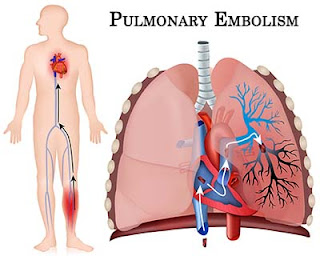
Brief Summary of Acute Pulmonary Embolism


Definition:

A pulmonary embolism results from the migration of venous thrombi from the systemic veins to pulmonary arterial system, resulting in varying degrees of obstruction of pulmonary arterial blood flow.
The incidence of pulmonary emboli in the United States exceeds 500,000 per year, with a mortality approaching 10%. If not diagnosed or if improperly treated, the mortality rate can reach 30%.

Common Sources:

Up to 90% of pulmonary emboli originate from the deep venous system of the legs. The upper extremities can also be a source of venous thrombi. Usually related to trauma, congenital fibromuscular bands, or the use of central venous catheters, 12% of all upper extremity thrombi result in pulmonary emboli. In addition, blood clot formation in the pelvic veins may cause either septic or bland pulmonary emboli, especially in the setting of complicated obstetric procedures or gynecologic surgery.
Other causes of pulmonary arterial obstructive emboli include air introduced during intravenous injections, hemodialysis, or the placement of central venous catheters; amniotic fluid secondary to vigorous uterine contractions; fat as a result of multiple long bone fractures; parasites; tumor cells; or injected foreign material (talc, mercury).

Risk Factors
:
Three basic risk factors, known collectively as Virchow’s triad, are associated with thrombus formation and subsequent pulmonary emboli:
  1. stasis,
  2. hypercoagulability, and 
  3. endothelial injury.

Management Of Acute Pulmonary Embolism


The goal of therapy is to prevent further embolic episodes, and heparin is the initial drug of choice for accomplishing this. First, a large intravenous loading bolus should be given, followed by continuous-drip infusion, maintained for at least 5 and often 7 to 10 days. Anticoagulation should not be withheld pending the results of further studies unless the patient’s risk of bleeding complications is greater than the clinical suspicion of pulmonary emboli. The partial thromboplastin time should be monitored and the heparin dosage adjusted to keep the time between 1.5 to 2.0 times the control.

Warfarin is started 24 to 48 hours after heparin therapy has been initiated. During the first 3 days of warfarin therapy, the prothrombin time or INR is increased before the onset of true anticoagulation. Therefore, before discontinuing the heparin, the prothrombin time or INR should be therapeutic (1.5 to 2 times normal) for approximately 2 to 3 days.

Low-molecular-weight heparins are indicated for prophylaxis in postoperative patients and probably have a role in the management of acute pulmonary embolism and deep venous thrombosis because they do not require monitoring of the anticoagulation effects.

Continuing the Treatment:
Long-term anticoagulation is usually achieved with warfarin, although low-molecular-weight heparins can also be used. Patients with reversible risk factors that are subsequently eliminated should undergo anticoagulation for a total of 3 months.

Sunday, May 21, 2017

Special considerations in Patients with Myocardial infarction


With myocardial infarction (MI), also known as heart attack, reduced blood flow through one of the coronary arteries results in myocardial ischemia and necrosis. With cardiovascular disease, the leading cause of death in the United States and western Europe, death usually results from the cardiac damage or complications of an MI.

Mortality is high when treatment is delayed; almost half of all sudden deaths due to an MI occur before hospitalization, within 1 hour of the onset of symptoms. The prognosis improves if vigorous treatment begins immediately.
  • Care for patients who have suffered an MI is directed toward detecting complications, preventing further myocardial damage, and promoting comfort, rest, and emotional well-being. Most MI patients receive treatment in the coronary care unit (CCU), where they’re under constant observation for complications.
  • On admission to the CCU, record the patient’s blood pressure, temperature, and heart and breath sounds, and monitor them regularly. Also, obtain an ECG.
  • Assess and record the severity and duration of pain; administer an analgesic. Avoid I.M. injections; absorption from the muscle is unpredictable.
  • Check the patient’s blood pressure after giving nitroglycerin, especially the first dose.
  • Frequently monitor the ECG to detect rate changes or arrhythmias. Place rhythm strips in the patient’s chart periodically for evaluation.
  • During episodes of chest pain, obtain ECG, blood pressure, and pulmonary artery catheter measurements for changes.
  • Watch for signs and symptoms of fluid retention (crackles, cough, tachypnea, and edema), which may indicate impending heart failure. Carefully monitor daily weight, intake and output, respirations, serum enzyme levels, and blood pressure.
  • Auscultate for adventitious breath sounds periodically (patients on bed rest frequently have atelectatic crackles, which may disappear after coughing) and for third or fourth heart sounds.
  • Organize patient care and activities to maximize periods of uninterrupted rest.

Tuesday, May 16, 2017

Hypertensive Problems - Approach In The Emergency Room


Bear the following points in mind when managing a hypertensive patient in the Emergency Department:
• Most patients with hypertension are asymptomatic.
• Hypertension is an important risk factor for cardiovascular disease and stroke.
• Most patients found to be hypertensive in the ED do not require any immediate intervention or treatment, but do require careful followup — usually by their GP.
• Never intervene on the basis of a single raised BP measurement in the absence of any associated symptoms and signs.

Approach
Approach patients found to be hypertensive as follows:
• Those with no previous history of hypertension, and no other concerns or history of other conditions (eg diabetes, peripheral vascular disease, IHD, or stroke) — arrange follow-up and monitoring with GP.
• Those known to be hypertensive already on treatment — arrange follow-up and monitoring with GP.
• Those displaying evidence of end organ damage (eg LV hypertrophy, retinal changes, renal impairment) — refer to the medical team.
• Those with hypertension associated with pain, vasoconstriction (eg acute pulmonary oedema) or stroke — treat underlying cause where possible. Do not intervene in stroke associated hypertension except under the direction of a neurologist or stroke specialist.
• Those with hypertension directly associated with symptoms or signs — contact the medical team and consider whether intervention is appropriate.

Saturday, May 13, 2017

Approach To A Patient Presenting With Chest Pain


Introduction:
Always take chest pain seriously. It may reflect life-threatening illness. Triage patients with chest pain as ‘urgent’ and ensure that they are seen within a few minutes. Ischaemic heart disease is understandably the first diagnosis to spring to mind in the middle-aged or elderly, but chest pain may have a variety of other disease processes, many of which are also potentially life-threatening.

The differential diagnosis of chest pain:

Common causes

  • Musculoskeletal (eg costochondritis)
  • Acute coronary syndrome
  • Pneumothorax
  • Oesophagitis
  • Pneumonia
  • Pulmonary embolism
Less common causes
  • Aortic dissection
  • Cholecystitis
  • Herpes zoster
  • Oesophageal rupture
  • Pancreatitis
  • Vertebral collapse

Reaching the correct conclusion requires accurate interpretation of the history, examination and investigations, bearing in mind recognized patterns of disease presentations.

History
Characterize the pain
• Site (eg central, bilateral or unilateral).
• Severity.
• Time of onset and duration.
• Character (eg ‘stabbing’, ‘tight/gripping’, or ‘dull/aching’).
• Radiation (eg to arms and neck in myocardial ischaemia).
• Precipitating and relieving factors (eg exercise/rest/GTN spray).
• Previous similar pains.

Wednesday, May 10, 2017

Electrocardiogram Interpretation - The Basics


Electrocardiogram interpretation

The Basics

  • The electrocardiogram (ECG) is normally recorded so that a deflection of 10mm = 1mV. 
  • The recording rate is 25mm/sec, 1mm = 0.04sec, 
  • 1 large square = 0.2sec. 

Rate Calculate the rate by dividing 300 by the number of large squares in one R–R interval.

Frontal plane axis Normally lies between –30 * and + 90 *. With a normal axis, QRS complexes in I and II are both + ve. An axis more –ve than –30 * (I + ve, aVF and II –ve) is left axis deviation (causes: left anterior hemiblock, inferior myocardial infarction (MI), ventricular tachycardia (VT), Wolf Parkinson White (WPW) syndrome). An axis more + ve than + 90 * (I –ve, aVF + ve) is right axis deviation (causes: pulmonary embolism (PE), cor pulmonale, lateral MI, left posterior hemiblock).


P wave Normally <0.12sec wide and <2.5mm tall. They are best seen in leads II and V 1 which are chosen for rhythm strips or monitoring.

  • A tall peaked P wave in II may reflect right atrial hypertrophy; 
  • a widened bifid P wave left atrial hypertrophy. 
  • P waves are absent in atrial fibrillation (AF).

PR interval Normally 0.12–0.2sec (<5 small squares).

  • A short PR interval (abnormally fast conduction between atria and ventricles) implies an accessory pathway (eg Wolf Parkinson White syndrome).
  • A prolonged PR interval occurs in heart block (first, second or third degree.

Friday, May 5, 2017

Abdominal Aneurysm


Abdominal aneurysm,
an abnormal dilation in the arterial wall generally occurs in the aorta between the renal arteries and iliac branches. 

Such aneurysms are four times more common in men than in women and are most prevalent in whites ages 50 to 80. More than 50% of all people with untreated abdominal aneurysms die within 2 years of diagnosis, primarily from rupture of the aneurysm; more than 85%, within 5 years.

Causes
About 95% of abdominal aortic aneurysms result from arteriosclerosis; the rest, from cystic medial necrosis, trauma, syphilis, and other infections. These aneurysms develop slowly.
First, a focal weakness in the muscular layer of the aorta (tunica media), due to degenerative changes, allows the inner layer (tunica intima) and outer layer (tunica adventitia) to stretch outward. Blood pressure within the aorta progressively weakens the vessel walls and enlarges the aneurysm.

Signs and symptoms
  • Although abdominal aneurysms usually don’t produce symptoms, most are evident (unless the patient is obese) as a pulsating mass in the periumbilical area, accompanied by a systolic bruit over the aorta. Some tenderness may be present on deep palpation. A large aneurysm may produce symptoms that mimic renal calculi, lumbar disk disease, and duodenal compression. Abdominal aneurysms rarely cause diminished peripheral pulses or claudication unless embolization occurs.
  • Pain, rupture, and hemorrhage
  • Lumbar pain that radiates to the flank and groin from pressure on lumbar nerves may signify enlargement and imminent rupture. If the aneurysm ruptures into the peritoneal cavity, it causes severe, persistent abdominal and back pain, mimicking renal or ureteral colic.
  • Signs and symptoms of hemorrhage—such as weakness, sweating, tachycardia, and hypotension—may be subtle because rupture into the retroperitoneal space produces a tamponade effect that prevents continued hemorrhage. Patients with such rupture may remain in stable condition for hours before shock and death occur, although 20% die immediately.

Wednesday, May 3, 2017

Arterial occlusive disease


Introduction: 
With arterial occlusive disease, the obstruction or narrowing of the lumen of the aorta and its major branches causes an interruption of blood flow, usually to the legs and feet. Arterial occlusive disease may affect the carotid, vertebral, innominate, subclavian, mesenteric, or celiac artery. Occlusions, which may be acute or chronic, often cause severe ischemia, skin ulceration, and gangrene.
Arterial occlusive disease is more common in males than in females. The prognosis depends on the location of the occlusion, the development of collateral circulation to counteract reduced blood flow and, if the patient has acute disease, the time elapsed between occlusion and its removal.
Causes
Arterial occlusive disease is a common complication of atherosclerosis. The occlusive mechanism may be endogenous, due to embolus formation or thrombosis, or exogenous, due to trauma or fracture. Predisposing factors include smoking; aging; conditions such as hypertension, hyperlipidemia, and diabetes; and a family history of vascular disorders, myocardial infarction, or stroke.
Signs and symptoms
Evidence of this disease varies widely, according to the occlusion site. (see table at the end of the article)

Diagnosis
With arterial occlusive disease, the diagnosis is usually based on patient history and physical examination.
Pertinent supportive diagnostic tests include the following:

Sunday, April 30, 2017

Managing Hypertensive Emergency


A hypertensive emergency (formerly called “malignant hypertension”) is severe hypertension (high blood pressure) with acute impairment of one or more organ systems (especially the central nervous system, cardiovascular system and/or the renal system) that can result in irreversible organ damage. In a hypertensive emergency, the blood pressure should be substantially lowered over a period of minutes to hours with an anti hypertensive agent.

Definition
The term hypertensive emergency is primarily used as a specific term for a hypertensive crisis with a diastolic blood pressure greater than or equal to 120 mmHg and/or systolic blood pressure greater than or equal to 180mmHg. Hypertensive emergency differs from hypertensive crisis in that, in the former, there is evidence of acute organ damage.

Laboratory Evaluation

Obtain electrolyte levels, as well as measurements of blood urea nitrogen (BUN) and creatinine levels to evaluate for renal impairment. A dipstick urinalysis to detect hematuria or proteinuria and microscopic urinalysis to detect red blood cells (RBCs) or RBC casts should also be performed

A complete blood cell (CBC) and peripheral blood smear should be obtained to exclude microangiopathic anemia, and a toxicology screen, pregnancy test, and endocrine testing may be obtained, as needed.

Management

If a patient presents to the emergency department with a high B.P the role of the treating physician is to determine either the patient is exhibiting any signs of end organ damage or not.

Thus, optimal control of hypertensive situations balances the benefits of immediate decreases in BP against the risk of a significant decrease in target organ perfusion. The emergency physician must be capable of appropriately evaluating patients with an elevated BP, correctly classifying the hypertension, determining the aggressiveness and timing of therapeutic interventions, and making appropraite decisions.

Acutely lowering the B.P in some situations may have an adverse effect too.

Pharmacological measures
An important point to remember in the management of the patient with any degree of BP elevation is to “treat the patient and not the number.” In patients presenting with hypertensive emergencies, antihypertensive drug therapy has been shown to be effective in acutely decreasing blood pressure.

Mitral Valve Prolapse


Mitral valve bulging back into the left atrium during systole is called mitral valve prolapse. This is also called floppy mitral valve or myxomatous mitral valve.

Etiology
MVP exhibits a strong hereditary component and in some patients is transmitted as an autosomal dominant trait.

Second common cause is rheumatic fever mostly seen in developing countries.

It is twice as frequent in women as in men.

MVP may occasionally result from Marfan syndrome, Ehler danlos syndrome and osteogenesis imperfecta.

Pathology
It develops due to myxomatous degeneration of mitral valve in which middle layer of valve leaflet composed of loose, myxomatous material is unusually prominent.

Clinical Features
Symptoms
  • Most patients with mitral valve prolapse are asymptomatic.
  • In symptomatic patients it may present as chest pain, dyspnea, fatigue, palpitation, syncopeand even sudden death.
  • Spontaneous rupture of chordea tendinae may cause a sudden worsening of MR that is hemodynamically severe.

Wednesday, April 26, 2017

Clinical Features Of Patients Presenting With Angina


Definition: 
Angina is defined as a condition in which patient suffers from chest pain due to decreased blood supply to the cardiac muscles. Most common cause is obstruction or spasm of coronary arteries.

Clinical Presentation
Diagnosis is usually made on clinical history. Patients with stable angina present with:

Pain: Episodic central-crushing or band like chest pain that may radiate to jaw/ neck or one/both arms. Pain in the arm/ neck may be the only symptom. It is important to ask about the frequency, severity, duration and timings of the pain.

Precipitating/ Relieving Factors: Angina pain is usually precipitated by exertion, cold, emotion, and/or heavy meals. Pain stops with rest or using glyceral trinitrate.

Associated Symptoms:  May be associated with palpitations, sweating, nausea and /or breathlessness during attacks.

Presence of risk factors: Patients who suffer from angina have certain risk factors like smoking, family history of cardiac disease, history of other vascular disease.

Monday, April 24, 2017

Managing High Blood Pressure


Introduction:

Millions of people around the globe have been diagnosed with high blood pressure or hypertension and it is one of the most common risk factor for stroke and heart attack. High blood pressure can also lead to damage to the blood vessels leading to kidney problems, aneurysms as well as damage to the eyes. It is very important to prevent, control and treat hypertension appropriately.

Guidelines For Target Blood Pressure:

The new guidelines recommend the following target for controlling high blood pressure:
  • among adults age 60 and older with high blood pressure, aim for a target blood pressure under 150/90.
  • among adults age 30 to 59 with high blood pressure, aim for a target blood pressure under 140/90
  • among adults with diabetes or chronic kidney disease, aim for a target blood pressure under 130/80.
Management:Managing high blood pressure involves diet changes, exercise, life style changes and if requires certain medications.

1. Loosing extra weight: Blood pressure often increases as weight increases. Loosing just 10 pounds helps reduces the blood pressure. Beside just loosing weight it is important to keep an eye on the waistline since carrying too much weight around the waistline leads to an increase in blood pressure.

2. Regular Exercise: Exercising regularly has a great impact on preventing and controlling high blood pressure. Even moderate activity for 10 minutes at a time or just walking may alos help a lot.

3. Eating A healthy diet: Aim to eat a diet rich in fruits, vegetables, whole grains and high fiber food. avoid fatty foods and also limit sugar intake.

4. Reduce Sodium In the diet: Even a small reduction of sodium in the diet can help reduce blood pressure greatly. Avoid eating processed food like chips and fast food items that are high in sodium content.

Angina And Its Treatment


Angina is a pain that comes from the heart usually as a result of narrowing of the coronary arteries and as a result the heart muscle does not get as much blood and oxygen supply as needed. The pain of angina is described as an uncomfortable pressure, discomfort and squeezing in the center of the chest. Sometimes the pain may be felt in the arm, neck, jaw or shoulder. A number of medications and treatment options are available for managing angina.

Non-Pharmacological Treatment:
  • If the patient smokes it is important to make every possible effort to stop him smoking.
  • The blood pressure should be checked regularly and if high should be manged accordingly.
  • If the patient is over weight , loosing weight is advised.
  • Avoid inactivity and the aim should be a moderate physical activity on most of the days of the week for at least 30 minutes daily.
  • If blood cholesterol is high it should be treated
  • The patient should eat a healthy diet and avoid fatty foods.
  • Avoid drinking alcohol.
  • Avoid taking large heavy meals that may trigger an attack of angina.
  • Patients are advised to slow down or take rest if physical activity triggers an attack of angina.
  • Some patients may get an angina due to a emotional stress so try to avoid the situations that causes stress.
Pharmacological Treatment:

1. Nitrates: are the most common drugs used for the angina treatment. Among the nitrates Nitroglycerin is the most commonly used medicine. Nitrates act by relaxing and dilating the blood vessels. This allows more blood to flow to the heart and also decreases the work load of the heart. Nitroglycerin or glyceryl trinitrate medicine comes in the form of a tablet or a spray and the dose is given under the tongue when the pain of angina develops. The medicine is absorbed quickly into the blood stream and acts within minutes to relieve the pain. The patients with a history of angina or those who are at risk are advised to carry the medicine with them all the time and use it as soon as needed. The most common side effect with this drug is a headache which usually improves with continued use.

Sunday, April 23, 2017

Introduction To Atrial Fibrillation


Atrial Fibrillation is the most common irregular heart rhythm and is characterized by irregular, disorganized electrical signals that cause the upper two chambers of the heart -the atria to contract very fast and irregularly.

The Physiology:
In order to understand atrial fibrillation it is important to explain the normal physiology of the heart’s electrical system. Each heart beat occurs due to the spread of the electrical signals that originate in the sino-atrial node located in the upper part of the right atrium, from where it travels to the right and the left atria and then to the atrio-ventricular node. After a slight delay the signals then pass to the ventricles.

In atrial fibrillation the signals do not generate in the sino-atrial node , instead they arise from another part of the atria and they do not travel normally. Theses abnormal signals spread through out the atria in a rapid disorganised way causing the atria to fibrillate meaning to contract fast and irregularly. Because of this faulty spread of the signals the atria and ventricles do not contract in a coordinated way , creating a fast , irregular heart rhythm.

The heart rate may reach up to 100 to 175 beats per minute in contrast to a normal heart rate of 60 to 100 beats per minute, and the ventricles cannot pump the blood to the body in a normal pattern.

Causes And Risk factors For Atrial Fibrillation:Damage to the heart’s electrical system causes atrial fibrillation. There are several conditions that may lead to atrial fibrillation like:

Deep Vein Thrombosis


Introduction:

Deep vein thrombosis is a condition in which a blood clot forms in one or more of the deep veins usually in the legs. A potentially life threatening complication may occur if this clot detaches and travels to the lungs leading to pulmonary embolism.

Etiology:
DVT (Deep vein thrombosis) results from conditions that impair venous return, lead to endothelial injury or dysfunction, or cause hypercoagulability. The conditions leading to DVT and the risk factors include:
  • Occult cancer.
  • Immobilized patients.
  • Adults over the age of 60 years
  • Recent history of a major surgery or trauma
  • Prolonged sitting like long travels
  • Family history of blood clots.
  • Pregnancy or delivery in last 6 months
  • Obesity
  • Fracture of the pelvis or legs.
  • Taking oral contraceptives or hormonal replacement therapy.
  • Certain autoimmune disorders
  • Cigarette smoking.
Clinical Signs And Symptoms:The clinical features of deep vein thrombosis include:
  • Swelling in the affected leg. ( Rarely both legs are involved)
  • Changes in skin color
  • Pain in the leg
  • Warmth and tenderness over the affected part
  • Positive Homan’s sign ( Calf discomfort elicited by ankle dorsiflexion with the knee extended)

Cardiac Tumors - A Brief Introduction


Most cardiac tumors are secondary deposits. Cardiac metastasis occurs most commonly with lung and breast carcinomas. The most commonprimary cardiac tumor is a myxoma; 75% of myxomas occur in the left atrium, the remainder in the right atrium and ventricle. Myxomas usually arise from the endocardium at the border of the fossa ovalis as a pedunculated mass. This may prolapse through the mitral valve (MV) mimicking mitral stenosis.

Epidemiology:
The prevalence of myxoma is estimated at 2 per 100,000, most commonly in those aged 30–60 years. The female to male ratio is 2:1.

Clinical presentation:
Myxomas are discovered when individuals present withconstitutional upset or the effects of MV obstruction, or the tumour is an incidental finding. Symptoms include
  • fever,
  • malaise,
  • exertional
  • dyspnoea and
  • weight loss.
  • Transient
  • pulmonary oedema,
  • paroxysmal
  • nocturnal dyspnoea,
  • haemoptysis,
  • dizziness and syncope may occur.
  • The first presentation may be due to an embolic phenomenon.

Aortic Dissection - An Emergency Medical Condition

Introduction: Aortic Dissection is a condition in which the aortic intima tears, exposing a diseased media that is split in two longitudinally by the force of the blood flow. This dissection usually progresses distally for a variable distance. Medial degeneration is often idiopathic but may be the result of cystic medial necrosis, especially in Marfan’s syndrome.

Associations: It is commonly seen in patients with Marfan syndrome. There is also an association with the following:
• hypertension (history of hypertension in 80% of cases);
• pregnancy;
• trauma.

Disease Classification: Aortic dissection is classified according to whether there is involvement of the ascending aorta (Stanford classification). This has practical and prognostic implications.


Epidemiology:
• Peak age 60 years
• Male/female ratio 2:1.

Clinical Presentation:
1.Common: There is central chest pain in 90% of cases, classically a ‘tearing’ pain that migrates to