Showing posts with label Emergency Medicine. Show all posts

Tuesday, August 24, 2021

Management Of Minor Burns


Minor burns can occur anytime while working in the kitchen or accidents can happen with young kids. Immediate first aid management of burns is important to know . 

First degree minor burns are usually not life threatening but are very painful and may even leave a scar if not properly treated.  

The skin is red and painful and there may be some swelling. 

1. First of all remove the victim and move to a safe place to avoid further burning. 

2. Cool the hot burned skin under cool running water or immerse in cool water for sometime , this helps to relieve the pain. 

3. Apply a moisturizing lotion like the one with aleo vera.

4. You can apply petroleum jelly 2-3 times a day. Avoid putting butter, too much of oil or lotions with fragrances which may cause an infection. 

5. Cover the burned skin with a sterile non sticking bandage .

6. If blisters form never pop them up. 

7. Take an over the counter pain killer like paracetamol or ibuprofen.

8. Protect the burned area from sun exposure.

9 If any signs of infection antibiotics may be needed

Thursday, July 20, 2017

Shock - A Brief Discussion


Shock is a condition of severe impairment of tissue perfusion leading to cellular injury and dysfunction. Rapid recognition and treatment are essential to prevent irreversible organ damage and death.

Different Causes And Categories Of Shock:

Hypovolemic shock

  • Hemorrhage
  • Intravascular volume depletion (e.g., vomiting, diarrhea, ketoacidosis)
  • Internal sequestration (ascites, pancreatitis, intestinal obstruction)

Cardiogenic shock

  • Myopathic (acute MI, fulminant myocarditis)
  • Mechanical (e.g., acute mitral regurgitation, ventricular septal defect, severe aortic stenosis, aortic dissection with aortic insufficiency)
  • Arrhythmic

Extracardiac obstructive shock

  • Pericardial tamponade
  • Massive pulmonary embolism
  • Tension pneumothorax

Distributive shock (profound decrease in systemic vascular tone)

  • Sepsis
  • Toxic overdoses
  • Anaphylaxis
  • Neurogenic (e.g., spinal cord injury)
  • Endocrinologic (Addison’s disease, myxedema)
Clinical Manifestations:

• Hypotension (mean arterial BP <60 mmHg), tachycardia, tachypnea, pallor, restlessness, and altered sensorium.
• Signs of intense peripheral vasoconstriction, with weak pulses and cold clammy extremities. In distributive (e.g., septic) shock, vasodilation predominates and extremities are warm.
• Oliguria (<20 mL/h) and metabolic acidosis common.
• Acute lung injury and acute respiratory distress syndrome with noncardiogenic pulmonary edema, hypoxemia, and diffuse pulmonary infiltrates.

Approach To The Patient: 

Obtain history for underlying causes, including 
  • cardiac disease (coronary disease, heart failure, pericardial disease), 
  • recent fever or infection leading to sepsis, 
  • drug effects (e.g., excess diuretics or ), 
  • conditions leading to pulmonary embolism and 
  • potential sources of bleeding.

Approach To A Patient With Chronic Obstructive Pulmonary Disease (COPD)


Chronic obstructive pulmonary disease (COPD) is characterized by chronic airflow limitation due to impedance to expiratory airflow, mucosal edema, infection, bronchospasm and bronchoconstriction due to decreased lung elasticity.

Smoking is the main cause, but others are chronic asthma, α-1 antitrypsin deficiency and chronic infection (eg bronchiectasis).

History
Exertional dyspnoea, cough, and sputum are usual complaints.
Ask about:
• Present treatment including inhalers, steroids, antibiotics, theophyllines, nebulizers, opiate analgesia, and home O 2 treatment.
• Past history: inquire about previous admissions and co-morbidity.
• Exercise tolerance: how far can they walk on the flat without stopping? How many stairs can they climb? Do they get out of the house?
• Recent history: ask about wheeze and dyspnoea, sputum volume and color. Chest injuries, abdominal problems and other infections may cause respiratory decompensation.
• Read the hospital notes: have there been prior ICU assessments? Has the respiratory consultant advised whether ICU would be appropriate?

Examination

  • Examine for dyspnoea, tachypnoea, accessory muscle use, and lip-pursing.
  • Look for hyperinflation (‘barrel chest’) and listen for wheeze or coarse crackles (large airway secretions). 
  • Cyanosis, plethora (due to secondary polycythaemia) and right heart failure (cor pulmonale) suggest advanced disease. 
  • Look for evidence of hypercarbia: tremor, bounding pulses, peripheral vasodilatation, drowsiness, or confusion.
  • Check for evidence of other causes of acute dyspnoea, particularly: asthma, pulmonary edema, pneumothorax , Pulmonary embolism. Remember that these conditions may co-exist with COPD.

Saturday, July 8, 2017

Acute Asthma - Management


Acute severe asthma
(also referred to in Latin as status asthmaticus, or asthmatic status) is an acute exacerbation of asthma that does not respond to standard treatments of bronchodilators (inhalers) and steroids.
Initial treatment
Follow the steps as summarized below:

• Provide high flow O 2 .
• Put the trolley back and side rails up so the patient is sitting up and holding on to the side rails (to use pectoral muscles as accessory muscles of respiration).
• If the patient cannot talk, start treatment, but get senior Emergency department and ICU help in case intubation and ventilation are required.
• Check trachea and chest signs for pneumothorax.
• Ask about previous admissions to ICU.
• Administer high dose nebulized B 2 agonist (eg salbutamol 5mg or terbutaline 10mg), or 10 puffs of salbutamol into spacer device and face mask.
For severe asthma or asthma that rseponds poorly to the initial nebulizer, consider continuous nebulization.
• Give a corticosteroid: either prednisolone 40–50mg PO or hydrocortisone (preferably as sodium succinate) 100mg IV.
• Add nebulized ipratropium bromide (500mcg) to B 2 agonist treatment for patients with acute severe or life-threatening asthma or those with a poor initial response to B 2 agonist therapy.
• Consider a single dose of IV magnesium sulphate (1.2–2g IVI over 20min) after consultation with senior medical staff, for patients with acute severe asthma without a good initial response to inhaled bronchodilator therapy or for those with life-threatening or near-fatal asthma.
• Use IV aminophylline only after consultation with senior medical staff.
Some individual patients with near-fatal or life-threatening asthma with a poor response to initial therapy may gain additional benefit. The loading dose of IVI aminophylline is 5mg/kg over 20min unless on maintenance therapy, in which case check blood theophylline level and start IVI of aminophylline at 0.5–0.7mg/kg/hr.
• IV salbutamol is an alternative in severe asthma, after consultation with senior staff. Draw up 5mg salbutamol into 500mL 5 % dextrose and run at a rate of 30–60mL/hr.
• A patient who cannot talk will be unable to drink fluids and may be dehydrated.
• Avoid ‘routine’ antibiotics.
• Repeat ABG within an hour.
• Hypokalaemia may be caused or exacerbated by B 2 agonist and/or steroid therapy.

Sunday, June 25, 2017

Acute Asthma - Assessment


Make an initial assessment of the severity of acute asthma based upon a combination of

  • clinical features, 
  • peak flow measurement and 
  • pulse oximetry 

This is as outlined as below.

Moderate exacerbation of asthma
• Increasing symptoms.
• Peak fl ow 50–75 % best or predicted.
• No features of acute severe asthma (below).

Acute severe asthma
Any one of the following:
• Inability to complete sentences in 1 breath.
• Respiratory rate ≥25/min.
• Heart rate ≥110/min.
• Peak flow 33–50 % best or predicted.

Monday, June 12, 2017

Approach to A Patient Presenting With Difficulty In Breathing ( Dyspnea)


The normal adult respiratory rate is 11–18/min, with a tidal volume of 400–800mL. Acute dyspnoea That is difficult or labored breathing is a common presenting symptom.

Common causes of Acute Dyspnoea
Cardiac
• Cardiogenic pulmonary oedema.
• MI .
• Pulmonary embolism.
• Arrhythmias .
Respiratory
• Asthma  or exacerbation of COPD
• Pneumonia .
• Pleural effusion .
• Pneumothorax
Trauma
• Aspiration of foreign body or vomit .
• Pneumothorax/haemothorax .
• Flail chest .
• Drowning incident .
Others
• Hypovolaemia or fever from any cause.
• Hyperventilation syndrome .
• Respiratory compensation for metabolic acidosis (DKA, salicylate overdose).
Psychological
• Anxiety
• Panic attack

Tuesday, May 23, 2017

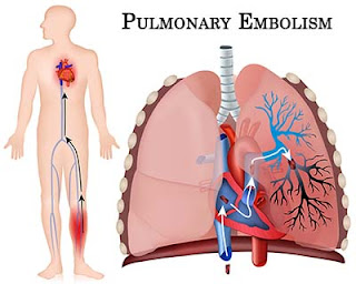
Management Of Acute Pulmonary Embolism


The goal of therapy is to prevent further embolic episodes, and heparin is the initial drug of choice for accomplishing this. First, a large intravenous loading bolus should be given, followed by continuous-drip infusion, maintained for at least 5 and often 7 to 10 days. Anticoagulation should not be withheld pending the results of further studies unless the patient’s risk of bleeding complications is greater than the clinical suspicion of pulmonary emboli. The partial thromboplastin time should be monitored and the heparin dosage adjusted to keep the time between 1.5 to 2.0 times the control.

Warfarin is started 24 to 48 hours after heparin therapy has been initiated. During the first 3 days of warfarin therapy, the prothrombin time or INR is increased before the onset of true anticoagulation. Therefore, before discontinuing the heparin, the prothrombin time or INR should be therapeutic (1.5 to 2 times normal) for approximately 2 to 3 days.

Low-molecular-weight heparins are indicated for prophylaxis in postoperative patients and probably have a role in the management of acute pulmonary embolism and deep venous thrombosis because they do not require monitoring of the anticoagulation effects.

Continuing the Treatment:
Long-term anticoagulation is usually achieved with warfarin, although low-molecular-weight heparins can also be used. Patients with reversible risk factors that are subsequently eliminated should undergo anticoagulation for a total of 3 months.

Sunday, May 21, 2017

Approach To A Patient Presenting With Haemoptysis (Blood In Sputum)


Haemoptysis (Blood In Sputum) may be the chief or sole complaint of patients presenting to the emergency department or their family doctors. . It always warrants investigation.

Causes of haemoptysis:

Respiratory causes;
• Infection (URTI, pneumonia, TB, lung abscess)
• Carcinoma (bronchial or laryngeal)
• Bronchiectasis

Cardiovascular causes:
• Pulmonary oedema
• PE
• Ruptured aortic aneurysm (aorto-bronchial fistula)

Coagulation disorder:
• Drugs (eg warfarin, heparin)
• Inherited (eg Haemophilia, Christmas disease)

Trauma:
• Penetrating or blunt

Others:
• Goodpasture’s, Wegener’s granulomatosis

Thursday, May 18, 2017

Mallory-Weiss syndrome


Mild to massive and usually painless bleeding due to a tear in the mucosa or submucosa of the cardia or lower esophagus characterizes Mallory-Weiss syndrome. Such a tear, usually singular and longitudinal, results from prolonged or forceful vomiting. About 60% of these tears involve the cardia; 15%, the terminal esophagus; and 25%, the region across the esophagogastric junction. Mallory-Weiss syndrome is most common in men older than age 40, especially alcoholics.

Causes
The direct cause of a tear in Mallory-Weiss syndrome is forceful or prolonged vomiting, probably when the upper esophageal sphincter fails to relax during vomiting. This lack of sphincter coordination is more common after excessive intake of alcohol. Other factors and conditions that may also increase intra-abdominal pressure and predispose to esophageal tearing include coughing, straining during bowel movements, trauma, seizures, childbirth, hiatal hernia, esophagitis, gastritis, and atrophic gastric mucosa.

Signs and symptoms
Typically, Mallory-Weiss syndrome begins with the vomiting of blood or the passing of large amounts of blood rectally a few hours to several days after normal vomiting. This bleeding, which may be accompanied by epigastric or back pain, may range from mild to massive but is generally more profuse than in esophageal rupture.
In patients with Mallory-Weiss syndrome, the blood vessels are only partially severed, preventing retraction and closure of the lumen. Massive bleeding—most likely when the tear is on the gastric side, near the cardia—may quickly lead to fatal shock.

Tuesday, May 16, 2017

Hypertensive Problems - Approach In The Emergency Room


Bear the following points in mind when managing a hypertensive patient in the Emergency Department:
• Most patients with hypertension are asymptomatic.
• Hypertension is an important risk factor for cardiovascular disease and stroke.
• Most patients found to be hypertensive in the ED do not require any immediate intervention or treatment, but do require careful followup — usually by their GP.
• Never intervene on the basis of a single raised BP measurement in the absence of any associated symptoms and signs.

Approach
Approach patients found to be hypertensive as follows:
• Those with no previous history of hypertension, and no other concerns or history of other conditions (eg diabetes, peripheral vascular disease, IHD, or stroke) — arrange follow-up and monitoring with GP.
• Those known to be hypertensive already on treatment — arrange follow-up and monitoring with GP.
• Those displaying evidence of end organ damage (eg LV hypertrophy, retinal changes, renal impairment) — refer to the medical team.
• Those with hypertension associated with pain, vasoconstriction (eg acute pulmonary oedema) or stroke — treat underlying cause where possible. Do not intervene in stroke associated hypertension except under the direction of a neurologist or stroke specialist.
• Those with hypertension directly associated with symptoms or signs — contact the medical team and consider whether intervention is appropriate.

Hypovolemic Shock - A Brief Discussion


In hypovolemic shock, reduced intravascular blood volume causes circulatory dysfunction and inadequate tissue perfusion. Without sufficient blood or fluid replacement, hypovolemic shock syndrome may lead to irreversible cerebral and renal damage, cardiac arrest and, ultimately, death. Hypovolemic shock requires early recognition of signs and symptoms and prompt, aggressive treatment to improve the prognosis.

Causes

Hypovolemic shock usually results from acute blood loss—about one-fifth of the total volume. Such massive blood loss may result from GI bleeding, internal hemorrhage (hemothorax and hemoperitoneum), or external hemorrhage (accidental or surgical trauma) or from any condition that reduces circulating intravascular plasma volume or other body fluids such as in severe burns. Other underlying causes of hypovolemic shock include intestinal obstruction, peritonitis, acute pancreatitis, ascites and dehydration from excessive perspiration, severe diarrhea or protracted vomiting, diabetes insipidus, diuresis, and inadequate fluid intake.

Signs and symptoms

Hypovolemic shock produces a syndrome of hypotension with narrowing pulse pressure; decreased sensorium; tachycardia; rapid, shallow respirations; reduced urine output; and cold, pale, clammy skin. Metabolic acidosis with an accumulation of lactic acid develops as a result of tissue anoxia as cellular metabolism shifts from aerobic to anaerobic pathways. Disseminated intravascular coagulation (DIC) is a possible complication of hypovolemic shock.

Sunday, May 14, 2017

A Brief Discussion on Snakebites (Poisonous)


Poisonous snakebites
are most common during summer afternoons in grassy or rocky habitats. Poisonous snakebites are medical emergencies. With prompt, correct treatment, they need not be fatal.

Causes
The only poisonous snakes in the United States are pit vipers (Crotalidae) and coral snakes (Elapidae). Pit vipers include rattlesnakes, water moccasins (cottonmouths), and copperheads. They have a pitted depression between their eyes and nostrils and two fangs, ¾? to 1¼? (2 to 3 cm) long. Because fangs may break off or grow behind old ones, some snakes may have one, three, or four fangs.
Because coral snakes are nocturnal and placid, their bites are less common than pit viper bites; pit vipers are also nocturnal but are more active. The fangs of coral snakes are short but have teeth behind them. Coral snakes have distinctive red, black, and yellow bands (yellow bands always border red ones), tend to bite with a chewing motion, and may leave multiple fang marks, small lacerations, and much tissue destruction.

Signs and symptoms
Most snakebites happen on the arms and legs, below the elbow or knee. Bites to the head or trunk are most dangerous, but any bite into a blood vessel is dangerous, regardless of location.

Most pit viper bites that result in envenomation cause immediate and progressively severe pain and edema (the entire extremity may swell within a few hours), local elevation in skin temperature, fever, skin discoloration, petechiae, ecchymoses, blebs, blisters, bloody wound discharge, and local necrosis.
Because pit viper venom is neurotoxic, pit viper bites may cause local and facial numbness and tingling, fasciculation and twitching of skeletal muscles, seizures (especially in children), extreme anxiety, difficulty speaking, fainting, weakness, dizziness, excessive sweating, occasional paralysis, mild to severe respiratory distress, headache, blurred vision, marked thirst and, in severe envenomation, coma and death. Pit viper venom may also impair coagulation and cause hema-temesis, hematuria, melena, bleeding gums, and internal bleeding. Other symptoms of pit viper bites include tachycardia, lymphadenopathy, nausea, vomiting, diarrhea, hypotension, and shock.

Wednesday, May 10, 2017

Disseminated Intravascular Coagulation


Introduction
Also called consumption coagulopathy and defibrination syndrome, disseminated intravascular coagulation (DIC) occurs as a complication of diseases and conditions that accelerate clotting. This accelerated clotting process causes small blood vessel occlusion, organ necrosis, depletion of circulating clotting factors and platelets, and activation of the fibrinolytic system—which, in turn, can provoke severe hemorrhage.
Clotting in the microcirculation usually affects the kidneys and extremities but may occur in the brain, lungs, pituitary and adrenal glands, and GI mucosa. Other conditions, such as vitamin K deficiency, hepatic disease, and anticoagulant therapy, may cause a similar hemorrhage.
DIC is generally an acute condition but may be chronic in cancer patients. The prognosis depends on early detection and treatment, the severity of the hemorrhage, and treatment of the underlying disease or condition.

Etiology

DIC may result from:
  • infection (the most common cause of DIC), including gram-negative or gram-positive septicemia; viral, fungal, or rickettsial infection; and protozoal infection (falciparum malaria)
  • obstetric complications, such as abruptio placentae, amniotic fluid embolism, and retained dead fetus
  • neoplastic disease, including acute leukemia and metastatic carcinoma
  • disorders that produce necrosis, such as extensive burns and trauma, brain tissue destruction, transplant rejection, and hepatic necrosis.
Other causes include heatstroke, shock, poisonous snakebite, cirrhosis, fat embolism, incompatible blood transfusion, cardiac arrest, surgery necessitating cardiopulmonary bypass, giant hemangioma, severe venous thrombosis, and purpura fulminans.
It isn’t clear why such disorders lead to DIC; nor is it certain that they lead to it through a common mechanism. In many patients, the triggering mechanisms may be the entrance of foreign protein into the circulation and vascular endothelial injury.

Management Of Patient Presenting With Electric Shock


When an electric current passes through the body, the damage it does depends on the intensity of the current (amperes, milliamperes, or microamperes), the resistance of the tissues it passes through, the kind of current (alternating current, direct current, or mixed), and the frequency and duration of current flow.
Electric shock may cause ventricular fibrillation, respiratory paralysis, burns, and death. The prognosis depends on the site and extent of damage, the patient’s state of health, and the speed and adequacy of treatment.

Causes
Electric shock usually follows accidental contact with exposed parts of electrical appliances or wiring, but it may also result from lightning or the flash of electric arcs from high-voltage power lines or machines.
The increased use of electrical medical devices in the hospital, many of which are connected directly to the patient, has raised serious concern for electrical safety and has led to the development of electrical safety standards. However, even well-designed equipment with reliable safety features can cause electric shock if it’s mishandled.
Signs and symptoms
Severe electric shock usually causes muscle contraction, followed by unconsciousness and loss of reflex control, sometimes with respiratory paralysis (by way of prolonged contraction of respiratory muscles or a direct effect on the respiratory nerve center).
After momentary shock, hyperventilation may follow muscle contraction. Passage of even the smallest electric current—if it passes through the heart— may induce ventricular fibrillation or another arrhythmia that progresses to fibrillation or myocardial infarction.
Electric shock from a high-frequency current (which generates more heat in tissues than a low-frequency current) usually causes burns as well as local tissue coagulation and necrosis. Low-frequency currents can also cause serious burns if contact with the current is concentrated in a small area—for example, when a toddler bites into an electrical cord.
Contusions, fractures, and other injuries can result from violent muscle contractions or falls during the shock; later, the patient may develop renal shutdown. Residual hearing impairment, cataracts, and vision loss may persist after severe electric shock.

Tuesday, May 9, 2017

Introduction to Tetanus


Introduction:
Tetanus, also known as lockjaw, is an acute exotoxin-mediated infection caused by the anaerobic, spore-forming, gram-positive bacillus Clostridium tetani. Usually, such infection is systemic; less often, localized.
Tetanus is fatal in up to 60% of non immunized persons, usually within 10 days of onset. When symptoms develop within 3 days after exposure, the prognosis is poor.

Etiology: Normally, transmission is through a puncture wound that is contaminated by soil, dust, or animal excreta containing C. tetani, or by way of burns and minor wounds. After C. tetani enters the body, it causes local infection and tissue necrosis. It also produces toxins that then enter the bloodstream and lymphatics and eventually spread to central nervous system tissue.
Tetanus occurs worldwide, but it’s more prevalent in agricultural regions and developing countries that lack mass immunization programs. It’s one of the most common causes of neonatal deaths in developing countries, where neonates of unimmunized mothers are delivered under unsterile conditions. In such neonates, the unhealed umbilical cord is the portal of entry.
In America, about 75% of all cases occur between April and September.

Clinical Features: 
The incubation period varies from 3 to 4 weeks in mild tetanus to less than 2 days in severe tetanus. When symptoms occur within 3 days after injury, death is more likely. If tetanus remains localized, signs of onset are spasm and increased muscle tone near the wound.
If tetanus is generalized (systemic), indications include 
  • marked muscle hypertonicity, 
  • hyperactive deep tendon reflexes, 
  • tachycardia, 
  • profuse sweating, 
  • low-grade fever, and 
  • painful, involuntary muscle contractions: 
  • neck and facial muscles, especially cheek muscles—locked jaw (trismus) and a grotesque, grinning expression called risus sardonicus 
  • somatic muscles—arched-back rigidity (opisthotonos), boardlike abdominal rigidity 
  • intermittent tonic convulsions lasting several minutes, which may result in cyanosis and sudden death by asphyxiation.

Sunday, May 7, 2017

Special Considerations In Cases With Traumatic Amputation


Traumatic amputation involves the accidental loss of a body part, usually a finger, a toe, an arm, or a leg. In complete amputation, the member is totally severed; in partial amputation, some soft-tissue connection remains.
The prognosis has improved as a result of early improved emergency and critical care management, new surgical techniques, early rehabilitation, prosthesis fitting, and new prosthesis design. New limb reimplantation techniques have been moderately successful, but incomplete nerve regeneration remains a major limiting factor.

Causes
Traumatic amputations usually result directly from accidents at a factory or farm, or from power tools or motor vehicle accidents.

Diagnosis
Any patient with a traumatic amputation requires careful monitoring of vital signs as well as assessment for other traumatic injuries. If amputation involves more than just a finger or a toe, assessment of airway, breathing, and circulation is also required. Because profuse bleeding is likely, watch for signs of hypovolemic shock, and draw blood for hemoglobin level, hematocrit, and typing and crossmatching. If the patient has a partial amputation, check for pulses distal to the amputation.

Friday, May 5, 2017

Abdominal Aneurysm


Abdominal aneurysm,
an abnormal dilation in the arterial wall generally occurs in the aorta between the renal arteries and iliac branches. 

Such aneurysms are four times more common in men than in women and are most prevalent in whites ages 50 to 80. More than 50% of all people with untreated abdominal aneurysms die within 2 years of diagnosis, primarily from rupture of the aneurysm; more than 85%, within 5 years.

Causes
About 95% of abdominal aortic aneurysms result from arteriosclerosis; the rest, from cystic medial necrosis, trauma, syphilis, and other infections. These aneurysms develop slowly.
First, a focal weakness in the muscular layer of the aorta (tunica media), due to degenerative changes, allows the inner layer (tunica intima) and outer layer (tunica adventitia) to stretch outward. Blood pressure within the aorta progressively weakens the vessel walls and enlarges the aneurysm.

Signs and symptoms
  • Although abdominal aneurysms usually don’t produce symptoms, most are evident (unless the patient is obese) as a pulsating mass in the periumbilical area, accompanied by a systolic bruit over the aorta. Some tenderness may be present on deep palpation. A large aneurysm may produce symptoms that mimic renal calculi, lumbar disk disease, and duodenal compression. Abdominal aneurysms rarely cause diminished peripheral pulses or claudication unless embolization occurs.
  • Pain, rupture, and hemorrhage
  • Lumbar pain that radiates to the flank and groin from pressure on lumbar nerves may signify enlargement and imminent rupture. If the aneurysm ruptures into the peritoneal cavity, it causes severe, persistent abdominal and back pain, mimicking renal or ureteral colic.
  • Signs and symptoms of hemorrhage—such as weakness, sweating, tachycardia, and hypotension—may be subtle because rupture into the retroperitoneal space produces a tamponade effect that prevents continued hemorrhage. Patients with such rupture may remain in stable condition for hours before shock and death occur, although 20% die immediately.

Tuesday, May 2, 2017

Introduction to Burns


A major burn is a horrifying injury, necessitating painful treatment and a long period of rehabilitation. It’s often fatal or permanently disfiguring and incapacitating (emotionally and physically). In the United States, about 2.5 million people annually suffer burns. It’s the nation’s third leading cause of accidental death.

Causes
Thermal burns, the most common type, are caused by flame, flash, scald or contact with hot objects. Examples are residential fires, motor vehicle accidents, playing with matches, improperly stored gasoline, space heater or electrical malfunctions, or arson. Other causes include improper handling of firecrackers, scalding accidents, and kitchen accidents (such as a child climbing on top of a stove or grabbing a hot iron). Burns in children are sometimes traced to parental abuse.
Chemical burns result from the contact, ingestion, inhalation, or injection of acids, alkalis, or vesicants that cause tissue injury and necrosis. 
Electrical burns result from coagulation necrosis caused by intense heat; they usually occur after contact with faulty electrical wiring or high-voltage power lines or when electric cords are chewed (by young children). Friction or abrasion burns happen when the skin is rubbed harshly against a coarse surface. 
Sunburn, of course, follows excessive exposure to sunlight.
Signs and symptoms
Symptoms will vary depending on the degree of burn. Suspect burn injury when the patient presents with blisters, pain, peeling skin, red skin, edema, white or charred skin, or signs of shock. Suspect an airway burn if you see charred mouth, burned lips, burns on the head, neck, or face; wheezing, change in voice, difficulty breathing and coughing; singed nose hairs or eyebrows; or dark carbon-stained mucous.

Monday, May 1, 2017

Managing Head Injuries


Head injury
can be defined as any alteration in mental or physical functioning related to a blow to the head. Loss of consciousness does not need to occur.

Head injury refers to trauma of the head. This may or may not include injury to the brain.

Acute Management
In the setting of acute head injury, give priority to the immediate assessment and stabilization of the airway and circulation.

Following stabilization, direct attention to prevention of secondary injury. Keep mean arterial pressures above 90 mm Hg; arterial saturations should be greater than 90%. Urgent CT scanning is a priority.

Most head injuries are of a benign nature and require no treatment beyond analgesics and close monitoring for potential complications such as intracranial bleeding. If the brain has been severely damaged by trauma, neurosurgical evaluation may be useful.

Monitoring Intracranial Pressure
Since elevated intracranial pressure is an independent predictor of poor outcome. If the intracranial pressure rises above 20-25 mm Hg, intravenous mannitol, CSF drainage, and hyperventilation can be used. Hypertonic saline has also been used in lieu of mannitol to lower intracranial pressure.

Sunday, April 30, 2017

Managing Hypertensive Emergency


A hypertensive emergency (formerly called “malignant hypertension”) is severe hypertension (high blood pressure) with acute impairment of one or more organ systems (especially the central nervous system, cardiovascular system and/or the renal system) that can result in irreversible organ damage. In a hypertensive emergency, the blood pressure should be substantially lowered over a period of minutes to hours with an anti hypertensive agent.

Definition
The term hypertensive emergency is primarily used as a specific term for a hypertensive crisis with a diastolic blood pressure greater than or equal to 120 mmHg and/or systolic blood pressure greater than or equal to 180mmHg. Hypertensive emergency differs from hypertensive crisis in that, in the former, there is evidence of acute organ damage.

Laboratory Evaluation

Obtain electrolyte levels, as well as measurements of blood urea nitrogen (BUN) and creatinine levels to evaluate for renal impairment. A dipstick urinalysis to detect hematuria or proteinuria and microscopic urinalysis to detect red blood cells (RBCs) or RBC casts should also be performed

A complete blood cell (CBC) and peripheral blood smear should be obtained to exclude microangiopathic anemia, and a toxicology screen, pregnancy test, and endocrine testing may be obtained, as needed.

Management

If a patient presents to the emergency department with a high B.P the role of the treating physician is to determine either the patient is exhibiting any signs of end organ damage or not.

Thus, optimal control of hypertensive situations balances the benefits of immediate decreases in BP against the risk of a significant decrease in target organ perfusion. The emergency physician must be capable of appropriately evaluating patients with an elevated BP, correctly classifying the hypertension, determining the aggressiveness and timing of therapeutic interventions, and making appropraite decisions.

Acutely lowering the B.P in some situations may have an adverse effect too.

Pharmacological measures
An important point to remember in the management of the patient with any degree of BP elevation is to “treat the patient and not the number.” In patients presenting with hypertensive emergencies, antihypertensive drug therapy has been shown to be effective in acutely decreasing blood pressure.