Showing posts with label Neurology. Show all posts

Thursday, June 29, 2017

Approach to a patient with Syncope


Definition Of Syncope: Symptom of sudden transient loss of consciousness due to global cerebral hypoperfusion.

Causes Of Syncope: 

• Neurocardiogenic (a.k.a. vasovagal, ~20%; ): ↑ sympathetic tone → vigorous contraction of LV →
mechanoreceptors in LV trigger ↑ vagal tone  → ↓ HR (cardioinhibitory) and/or
↓ BP (vasodepressor) cough, deglutition, defecation, & micturition → ↑ vagal tone and thus can be precipitants related disorder:
carotid sinus hypersensitivity (exagg vagal resp to carotid massage)

• Orthostatic hypotension (10%)
hypovolemia/diuretics, deconditioning; vasodilat. (esp. if combined w/ chronotropes)
autonomic neuropathy [primary = Parkinson’s, Shy-Drager, Lewy body dementia, POTS (dysautonomia in the young); secondary =DM, EtOH, amyloidosis, CKD]

• Cardiovascular
Arrhythmia (15%)
Bradyarrhythmias: Sick sinus syndrome, high-grade AV block, chronotropes, PPM malfunction
Tachyarrhythmias: VT, SVT (syncope rare unless structural heart disease or WPW)
Mechanical (5%)
Endocardial/Valvular: AS, MS, PS, prosthetic valve thrombosis, myxoma
Myocardial: pump dysfxn from MI or outflow obstruction from HCMP (but usually VT)
Pericardial: tamponade
Vascular: PE, PHT, aortic dissection, ruptured AAA, subclavian steal

Monday, June 12, 2017

Lumbar Puncture


A lumbar puncture (also called a spinal tap) is a procedure to collect and look at the fluid (cerebrospinal fluid, or CSF) surrounding the brain and spinal cord. During a lumbar puncture, a needle is carefully inserted into the spinal canal low in the back (lumbar area). Samples of CSF are collected.
Evaluation of CSF is essential for the diagnosis of suspected meningeal infection, subarachnoid hemorrhage, leptomeningeal neoplastic disease, and noninfectious meningitis.

Relative contraindications to LP include 

  • local skin infection in the lumbar area, 
  • suspected spinal cord mass lesion, and 
  • a suspected intracranial mass lesion. 
Preparation: 

  • Any bleeding diathesis should also be corrected prior to performing LP to prevent the possible occurrence of an epidural hematoma. 
  • A functional platelet count >50,000/μL and an international normalized ratio (INR) <1.5 are advisable to perform LP safely.
  • In patients with an altered level of consciousness, focal neurologic deficits, or evidence of papilledema, an imaging study should generally be obtained prior to performing LP.
Technique: 

  • Proper positioning of the patient is essential; either the lateral decubitus or the sitting position can be used. 
  • Most routine LPs should be performed using the lateral decubitus position .The sitting position may be preferable in obese patients. 
  • With either position, the patient should be instructed to flex the spine as much as possible. 
  • In the lateral decubitus position, the patient assumes a fetal position with the knees flexed toward the abdomen; the shoulders and pelvis should be vertically aligned without forward or backward tilt. 
  • In the sitting position, the patient should bend over a bedside table with the head resting on folded arms.

Monday, June 5, 2017

Medical Issues Common In Children With Neurodisability


There are a number of medical problems which are more common in children with disability. These
include:

1. Gastro-esophageal reflux disease (GERD) :Gastro-oesophageal reflux (the non-forceful regurgitation of gastric contents into the esophagus) is more common in children with neuro-developmental problems for a number of reasons. 
Intra-abdominal pressure may be increased for structural reasons such as scoliosis, the lower esophageal sphincter may be functionally immature as a reflection of abnormal muscle tone elsewhere in the body, and difficulties in upright positioning may also exacerbate gastroesophageal reflux.
First line management is postural, with supportive upright seating and sleeping positioners, along with adjustment of feed consistency if required. In addition, pharmacological agents like Proton-pump inhibitors (e.g. omeprazole, lansoprazole, esomeprazole) and H2-receptor antagonists (e.g. ranitidine) are used to alter acid production. Dopamine receptor antagonists (e.g. domperidone) stimulate gastric emptying and small intestinal transit. They also help with GERD by enhancing the tone of the esophageal sphincter. In children with severe symptoms of GORD, surgical intervention such as a Nissen’s fundoplication may be considered.

2. Respiratory complications: Neurological disturbance in children with neurodisability impairs the ability of the child to protect their airway, leading to acute or chronic (‘silent’) aspiration and liability to chemical pneumonitis or secondary infection with anaerobic organisms.

Saturday, May 27, 2017

Introduction to Hydrocephalus


An excessive accumulation of cerebrospinal fluid (CSF) within the ventricular spaces of the brain, hydrocephalus occurs most commonly in neonates. It can also occur in adults as a result of injury or disease. In infants, hydrocephalus enlarges the head, and in both infants and adults, the resulting compression can damage brain tissue.
With early detection and surgical intervention, the prognosis improves but remains guarded. Even after surgery, such complications as developmental delay, impaired motor function, and vision loss can persist. Without surgery, the prognosis is poor: Mortality may result from increased intracranial pressure (ICP) in people of all ages; infants may also die prematurely of infection and malnutrition.

Causes
Hydrocephalus may result from an obstruction in CSF flow (noncommunicating hydrocephalus) or from faulty absorption of CSF (communicating hydrocephalus).

Noncommunicating hydrocephalus
In noncommunicating hydrocephalus, the obstruction occurs most commonly between the third and fourth ventricles, at the aqueduct of Sylvius, but it can also occur at the outlets of the fourth ventricle (foramina of Luschka and Magendie) or, rarely, at the foramen of Monro.
This obstruction may result from faulty fetal development, infection (syphilis, granulomatous diseases, meningitis), a tumor, a cerebral aneurysm, or a blood clot (after intracranial hemorrhage).

Communicating hydrocephalus

In communicating hydrocephalus, faulty absorption of CSF may result from surgery to repair a myelomeningocele, adhesions between meninges at the base of the brain, or meningeal hemorrhage. Rarely, a tumor in the choroid plexus causes overproduction of CSF, producing hydrocephalus.

Brief Summary of Cerebral Palsy


The most common cause of crippling in children, cerebral palsy is an umbrella term for a group of neuromuscular disorders resulting from prenatal, perinatal, or postnatal central nervous system (CNS) damage. Although nonprogressive, these disorders may become more obvious as an affected infant ages.

Types:
Three major types of cerebral palsy occur—
  1. spastic, 
  2. athetoid, and 
  3. ataxic
—sometimes in mixed forms. Motor impairment may be minimal (sometimes apparent only during physical activities such as running) or severely disabling. Associated defects—such as seizures, speech disorders, and mental retardation—are common.
The prognosis varies. With mild impairment, proper treatment may make a near-normal life possible.

Cerebral palsy is most common in premature infants (anoxia plays the greatest role in contributing to cerebral palsy) and in those who are small for their gestational age. Cerebral palsy is common in whites and is slightly more common in males than in females.

Friday, May 26, 2017

Introduction to Stroke


Stroke
is a sudden impairment of cerebral circulation in one or more blood vessels supplying the brain. Stroke interrupts or diminishes oxygen supply and commonly causes serious damage or necrosis in brain tissues.
The sooner circulation returns to normal after a stroke, the better the chances are for complete recovery. However, about half of those who survive a stroke remain permanently disabled and experience a recurrence within weeks, months, or years.

Incidence
Stroke is the third most common cause of death in the United States today and the most common cause of neurologic disability. It affects 500,000 people each year; half of them die as a result.

Causes
Factors that increase the risk of stroke include history of transient ischemic attacks (TIAs), atherosclerosis, hypertension, electrocardiogram changes, arrhythmias, rheumatic heart disease, diabetes mellitus, gout, postural hypotension, cardiac or myocardial enlargement, high serum triglyceride levels, lack of exercise, use of hormonal contraceptives, cigarette smoking, and family history of stroke.

The major causes of stroke are thrombosis, embolism, and hemorrhage.

Management Of Patients Presenting With Stroke


Treatment options vary, depending on the type of stroke the patient experiences.

Early medical diagnosis of the type of stroke coupled with new drug treatments can greatly reduce the long-term disability secondary to ischemia.
Surgery performed to improve cerebral circulation for patients with thrombotic or embolic stroke includes an endarterectomy (the removal of atherosclerotic plaque from the inner arterial wall) or a microvascular bypass (the surgical anastomosis of an extracranial vessel to an intracranial vessel).

Medications useful in treating stroke include:
  • alteplase (recombinant tissue plasminogen activator), effective in emergency treatment of embolic stroke
  • (Patients with embolic or thrombotic stroke who aren’t candidates for alteplase [3 to 6 hours poststroke] should receive aspirin or heparin.)
  • long-term use of aspirin or ticlopidine, used as antiplatelet agents to prevent recurrent stroke
  • anticoagulants (heparin, warfarin), which may be required to treat crescendo TIAs not responsive to antiplatelet drugs
  • antihypertensives, antiarrhythmics, and antidiabetics, which may be used to treat risk factors associated with recurrent stroke.
Early supportive therapy
Frequently assess neurologic status, using the National Institutes of Health (NIH) Stroke Scale to determine deficits.
  • If the patient has been treated with alteplase, monitor him for signs of hemorrhage.
  • Monitor blood pressure frequently; give labetalol for severe hypertension.
  • Use acetaminophen and hypothermia blankets to control fever.
  • Maintain a patent airway and oxygenation status; intubate and ventilate the patient as needed.
  • Monitor blood glucose levels.
  • Monitor electrocardiogram results, and treat arrhythmias as early as possible.
  • If the patient develops a headache, administer an analgesic.

Saturday, May 20, 2017

Introduction to Neurogenic bladder


Introduction
Also known as neuromuscular dysfunction of the lower urinary tract, neurologic bladder dysfunction, and neuropathic bladder, neurogenic bladder refers to all types of bladder dysfunction caused by an interruption of normal bladder innervation. Subsequent complications include urinary incontinence, residual urine retention, urinary tract infection, calculi formation, and renal failure. A neurogenic bladder can be spastic (hypertonic, reflex, or automatic), flaccid (hypotonic, atonic, nonreflex, or autonomous), or uncoordinated (dyssynergic).

Causes
At one time, neurogenic bladder was thought to result primarily from spinal cord injury; now, it appears to stem from a host of underlying conditions:
  • cerebral disorders, such as cerebrovascular accident, brain tumor (meningioma and glioma), Parkinson’s disease, multiple sclerosis, and dementia
  • spinal cord disease or trauma, such as spinal stenosis (causing cord compression) or arachnoiditis (causing adhesions between the membranes covering the cord), cervical spondylosis, myelopathies from hereditary or nutritional deficiencies and, rarely, tabes dorsalis
  • disorders of peripheral innervation, including autonomic neuropathies resulting from endocrine disturbances such as diabetes mellitus (most common)
  • metabolic disturbances, such as hypothyroidism, porphyria, or uremia (infrequent)
  • acute infectious diseases such as Guillain-Barré syndrome
  • heavy metal toxicity
  • chronic alcoholism
  • collagen diseases such as systemic lupus erythematosus
  • vascular diseases such as atherosclerosis
  • distant effects of cancer such as primary oat cell carcinoma of the lung
  • herpes zoster
  • sacral agenesis.
An upper motor neuron lesion (above S2 to S4) causes spastic neurogenic bladder, with spontaneous contractions of the detrusor muscles, elevated intravesical voiding pressure, bladder wall hypertrophy with trabeculation, and urinary sphincter spasms.

A lower motor neuron lesion (below S2 to S4) causes flaccid neurogenic bladder, with decreased intravesical pressure, increased bladder capacity and large residual urine retention, and poor detrusor contraction.

Tuesday, May 16, 2017

Introduction to Bell’s palsy


Bell’s palsy
is a neurologic disorder that affects the seventh cranial (facial) nerve, producing unilateral facial weakness or paralysis. Onset is rapid. Although it affects all age-groups, it’s most common in persons younger than age 60. In 80% to 90% of patients, it subsides spontaneously, with complete recovery in 1 to 8 weeks; however, recovery may be delayed in older adults. If recovery is partial, contractures may develop on the paralyzed side of the face. Bell’s palsy may recur on the same or opposite side of the face.

Pathophysiology
The seventh cranial nerve is responsible for motor innervation of the facial muscles. With Bell’s palsy, the nerve is blocked by an inflammatory reaction around the nerve (usually at the internal auditory meatus). This is commonly associated with infections (most likely herpes simplex) and can result from hemorrhage, tumor, meningitis, or local trauma.

Signs and symptoms
Bell’s palsy usually produces unilateral facial weakness, occasionally with aching pain around the angle of the jaw or behind the ear. On the weak side, the mouth droops (causing the patient to drool saliva from the corner of his mouth), and taste perception is distorted over the affected anterior portion of the tongue. In addition, the forehead appears smooth, and the patient’s ability to close his eye on the weak side is markedly impaired. When he tries to close this eye, it rolls upward (Bell’s phenomenon) and shows excessive tearing. The patient also has hypersensitivities to sound.
Although Bell’s phenomenon occurs in those without Bell’s palsy, it isn’t apparent because the eye closes completely and covers this eye motion. In Bell’s palsy, incomplete eye closure makes this upward motion obvious.

Thursday, May 11, 2017

An Introduction to Epilepsy


Seizure disorder, or epilepsy, is a condition of the brain characterized by a susceptibility to recurrent seizures (paroxysmal events associated with abnormal electrical discharges of neurons in the brain). Epilepsy is believed to affect 1% to 2% of the population. The prognosis is good if the patient with epilepsy adheres strictly to his prescribed treatment.

Causes
In about one-half of all epilepsy cases, the cause is unknown. Possible causes include:
  • birth trauma (inadequate oxygen supply to the brain, blood incompatibility, or hemorrhage)
  • perinatal infection
  • anoxia
  • infectious diseases (meningitis, encephalitis, or brain abscess)
  • ingestion of toxins (mercury, lead, or carbon monoxide)
  • brain tumors
  • inherited disorders or degenerative disease, such as phenylketonuria or tuberous sclerosis
  • head injury or trauma
  • metabolic disorders, such as hypoglycemia and hypoparathyroidism
  • stroke (hemorrhage, thrombosis, or embolism).
Signs and symptoms
The hallmark of epilepsy is recurring seizures, which can be classified as partial, generalized, status epilepticus, or unclassified (some patients may be affected by more than one type).

Partial seizures

Arising from a localized area of the brain, partial seizures cause focal symptoms. These seizures are classified by their effect on consciousness and whether they spread throughout the motor strip, causing a generalized seizure. When simple or complex seizures evolve to both sides of the brain, they’re termed secondary generalized seizures.

A simple partial seizure begins locally and generally doesn’t cause an alteration in consciousness. It isn’t uncommon for this type to present with sensory symptoms (lights flashing, smells, hearing hallucinations), autonomic symptoms (sweating, flushing, pupil dilation), and psychic symptoms (dream states, anger, fear). The seizure lasts for a few seconds and occurs without preceding or provoking events.
A complex partial seizure involves impairment in consciousness. Amnesia for the events that occur during and immediately after the seizure is a differentiating characteristic. During the seizure, the patient may follow simple commands. This type of partial seizure generally lasts for 1 to 3 minutes.

Tuesday, May 9, 2017

Carpal Tunnel Syndrome


The most common of the nerve entrapment syndromes, carpal tunnel syndrome results from compression of the median nerve at the wrist, within the carpal tunnel. This nerve passes through, along with blood vessels and flexor tendons, to the fingers and thumb. Compression neuropathy causes sensory and motor changes in the median distribution of the hand.
Carpal tunnel syndrome usually occurs in women between ages 30 and 60 and poses a serious occupational health problem. Assembly-line workers and packers, secretary-typists, and persons who repeatedly use poorly designed tools are most likely to develop this disorder. Any strenuous use of the hands—ustained grasping, twisting, or flexing—aggravates this condition.

Causes
The carpal tunnel is formed by the carpal bones and the transverse carpal ligament. Inflammation or fibrosis of the tendon sheaths that pass through the carpal tunnel can cause edema and compression of the median nerve.
Many conditions can cause the contents or structure of the carpal tunnel to swell and press the median nerve against the transverse carpal ligament. Such conditions include rheumatoid arthritis, flexor tenosynovitis (commonly associated with rheumatic disease), nerve compression, pregnancy, renal failure, menopause, diabetes mellitus, acromegaly, edema following Colles’ fracture, hypothyroidism, amyloidosis, myxedema, benign tumors, tuberculosis, and other granulomatous diseases. Another source of damage to the median nerve is dislocation or acute sprain of the wrist.

Signs and symptoms

The patient with carpal tunnel syndrome usually complains of weakness, pain, burning, numbness, or tingling in the involved hands. This paresthesia affects the thumb, forefinger, middle finger, and half of the fourth finger. The patient is unable to clench his hand into a fist. The nails may be atrophic; the skin, dry and shiny.
Because of vasodilation and venous stasis, symptoms are usually worse at night and in the morning. The pain may spread to the forearm and, in severe cases, as far as the shoulder. The patient can usually relieve such pain by shaking his hands vigorously or dangling his arms at his side.

Sunday, May 7, 2017

Amyotrophic lateral sclerosis


Commonly called Lou Gehrig disease, after the New York Yankee first baseman who died of this disorder, amyotrophic lateral sclerosis (ALS) is the most common motor neuron disease causing muscular atrophy. Other motor neuron diseases include progressive muscular atrophy and progressive bulbar palsy. Onset occurs between ages 40 and 70. A chronic, progressively debilitating disease, ALS is rapidly fatal.

Causes
The disease is progressive, with death resulting from respiratory paralysis (the median survival is 3 to 5 years), and it’s three times more common in men than in women. The exact cause of ALS is unknown, but 5% to 10% of ALS cases have a genetic component. In these cases, it’s an autosomal dominant trait that affects men and women equally.

ALS and other motor neuron diseases may result from:
  • a slow-acting virus
  • nutritional deficiency related to a disturbance in enzyme metabolism
  • metabolic interference in nucleic acid production by the nerve fibers
  • an autoimmune disorder that affects immune complexes in the renal glomerulus and basement membrane.
Precipitating factors for acute deterioration include trauma, viral infections, and physical exhaustion.

Signs and symptoms
Patients with ALS develop fasciculations, accompanied by atrophy and weakness, especially in the muscles of the forearms and the hands. Other signs include impaired speech; difficulty chewing, swallowing, and breathing, particularly if the brain stem is affected; and, occasionally, choking and excessive drooling.
Mental deterioration doesn’t usually occur, but patients may become depressed as a result of the disease. Progressive bulbar palsy may cause crying spells or inappropriate laughter.

Saturday, May 6, 2017

Guillain-Barré Syndrome


Also known as infectious polyneuritis, Landry-Guillain-Barré syndrome, and acute idiopathic polyneuritis, Guillain-Barré syndrome is an acute, rapidly progressive, and potentially fatal form of polyneuritis that causes muscle weakness and mild distal sensory loss.
This syndrome can occur at any age but is most common between ages 30 and 50; it affects both sexes equally. Recovery is spontaneous and complete in about 95% of patients, although mild motor or reflex deficits in the feet and legs may persist. The prognosis is best when symptoms clear between 15 and 20 days after onset.

Causes
The precise cause of Guillain-Barré syndrome is unknown, but it may be a cell-mediated immune response with an attack on peripheral nerves in response to a virus. The major pathologic effect is segmental demyelination of the peripheral nerves.
Because this syndrome causes inflammation and degenerative changes in both the posterior (sensory) and the anterior (motor) nerve roots, signs of sensory and motor losses occur simultaneously.

Predisposing factors
About 50% of patients with Guillain-Barré syndrome have a recent history of minor febrile illness, usually an upper respiratory tract infection or, less commonly, gastroenteritis. When infection precedes the onset of Guillain-Barré syndrome, signs of infection subside before neurologic features appear.
Other possible precipitating factors include surgery, rabies or swine influenza vaccination, viral illness, Hodgkin’s or some other malignant disease, and systemic lupus erythematosus.

Signs and symptoms
Muscle weakness, the major neurologic sign, usually appears in the legs first (ascending type) and then extends to the arms and facial nerves within 24 to 72 hours. Sometimes, muscle weakness develops in the arms first (descending type) or in the arms and legs simultaneously. In milder forms of the disease, muscle weakness may affect only the cranial nerves or may not occur.
Paresthesia, another common neurologic sign, sometimes precedes muscle weakness but vanishes quickly. Some patients with the disorder never develop this symptom.
Other clinical features include facial diplegia (possibly with ophthalmoplegia [ocular paralysis]), dysphagia or dysarthria and, less commonly, weakness of the muscles supplied by cranial nerve XI (spinal accessory nerve).
Muscle weakness develops so quickly that muscle atrophy doesn’t occur, but hypotonia and areflexia do. Stiffness and pain in the form of a severe “charley horse” occur in many cases.

Friday, May 5, 2017

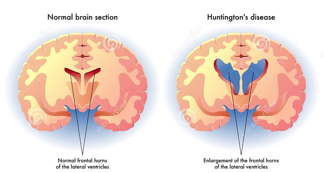
Huntington’s Disease


Also called Huntington’s chorea, hereditary chorea, chronic progressive chorea, and adult chorea, Huntington’s disease is a hereditary disease in which degeneration in the cerebral cortex and basal ganglia causes chronic progressive chorea (involuntary and irregular movements) and cognitive deterioration, ending in dementia.

Huntington’s disease usually strikes people between ages 25 and 55 (the average age is 35); however, 2% of cases occur in children, and 5%, as late as age 60. Males and females are equally affected. Death usually results 10 to 15 years after onset from suicide, heart failure, or pneumonia.

Causes
Huntington’s disease is transmitted as an autosomal dominant trait, and either sex can transmit and inherit it. Each child of a parent with this disease has a 50% chance of inheriting it; the child who inherits it can pass it on to his own children.
Because of hereditary transmission, Huntington’s disease is prevalent in areas in which affected families have lived for several generations. Genetic testing is offered to those with a known family history of the disease.
Signs and symptoms
The onset of this disease is insidious. The patient eventually becomes totally dependent—emotionally and physically—through loss of musculoskeletal control.
Neurologic manifestations
Gradually, the patient develops progressively severe choreic movements. Such movements are rapid, usually violent, and purposeless. Initially, they’re unilateral and more prominent in the face and arms than in the legs, progressing from mild fidgeting to grimacing, tongue smacking, dysarthria (indistinct speech), athetoid movements (slow, twisting muscle contractions, especially of the hands) related to emotional state, and torticollis (neck muscle contractions).
Bradykinesia (slow movements) is commonly accompanied by rigidity. Muscle strength is generally maintained. The combination of chorea, bradykinesia, and normal muscle strength results in impairment of both voluntary and involuntary movement.
Another neurologic manifestation is dysphagia, which is seen in a large percentage of patients in the advanced stages. Dysarthria generally presents early in the disease process and may be complicated by perseveration (persistent repetition of a reply), oral apraxia (difficulty coordinating movement of the mouth), and aprosody (inability to accurately reproduce or interpret the tone of language).

Thursday, May 4, 2017

Trigeminal Neuralgia


Introduction
Also called tic douloureux, trigeminal neuralgia is a painful disorder of one or more branches of the fifth cranial (trigeminal) nerve that produces paroxysmal attacks of excruciating facial pain precipitated by stimulation of a trigger zone.

It occurs mostly in people over age 40, in women more often than men, and on the right side of the face more often than the left. Trigeminal neuralgia can subside spontaneously, with remissions lasting from several months to years.
Etiology
Although the cause remains undetermined, trigeminal neuralgia may:
  • reflect an afferent reflex phenomenon located centrally in the brain stem or more peripherally in the sensory root of the trigeminal nerve
  • be related to compression of the nerve root by posterior fossa tumors, middle fossa tumors, or vascular lesions (subclinical aneurysm), although such lesions usually produce simultaneous loss of sensation
  • occasionally be a manifestation of multiple sclerosis or herpes zoster.
Whatever the cause, the pain of trigeminal neuralgia is probably produced by an interaction or short-circuiting of touch and pain fibers.
Clinical Features
Typically, the patient reports a searing or burning pain that occurs in lightning-like jabs and lasts from 1 to 15 minutes (usually 1 to 2 minutes) in an area innervated by one of the divisions of the trigeminal nerve, primarily the superior mandibular or maxillary division.
The pain rarely affects more than one division, and seldom the first division (ophthalmic) or both sides of the face. It affects the second (maxillary) and third (mandibular) divisions of the trigeminal nerve equally.

Wednesday, May 3, 2017

Tic disorders


Introduction
Including Tourette syndrome, chronic motor or vocal tic disorder, and transient tic disorder, tic disorders are similar pathophysiologically but differ in severity and prognosis. All tic disorders, commonly known simply as tics, are involuntary, spasmodic, recurrent, and purposeless motor movements or vocalizations. These disorders are classified as motor or vocal and as simple or complex.
Tics may begin as early as age 2, with average onset being age 7. All tic disorders are three times more common in boys than in girls. Transient tics usually are self-limiting, but Tourette syndrome follows a chronic course with remissions and exacerbations.
Causes
Although their exact cause is unknown, tic disorders occur more frequently in certain families, suggesting a genetic cause. Tics commonly develop when a child experiences overwhelming anxiety, usually associated with normal maturation. Tics may be precipitated or exacerbated by the use of phenothiazines or central nervous system stimulants or by head trauma.
Signs and symptoms
Assessment findings vary according to the type of tic disorder. 

Inspection, coupled with the patient’s history, may reveal the specific motor or vocal patterns that characterize the tic, as well as the frequency, complexity, and precipitating factors. The patient or his family may report that the tics occur sporadically many times a day.
Note whether certain situations exacerbate the tics. All tic disorders may be exacerbated by stress, and they usually diminish markedly during sleep. The patient also may report that they occur during activities that require concentration, such as reading or sewing.
Determine whether the patient can control the tics. Most patients can, with conscious effort, control them for short periods.

Monday, May 1, 2017

Parkinson’s Disease


Parkinson disease is recognized as one of the most common neurologic disorders, affecting approximately 1% of individuals older than 60 years.

Parkinson’s disease (also known as Parkinson disease, Parkinson’s, idiopathic parkinsonism, primary parkinsonism, PD, or paralysis agitans) is a degenerative disorder of the central nervous system. The motor symptoms of Parkinson’s disease result from the death of dopamine generating cells in the substantia nigra, a region of the midbrain.

The classic motor features of Parkinson disease typically start insidiously and emerge slowly over weeks or months, with tremor being the most common initial symptom. The 3 cardinal signs of Parkinson disease are resting tremor, rigidity, and bradykinesia.

Pathology
No specific, standard criteria exist for the neuropathologic diagnosis of Parkinson disease, as the specificity and sensitivity of its characteristic findings have not been clearly established. However, the following are the 2 major neuropathologic findings in Parkinson disease:
Loss of pigmented dopaminergic neurons of the substantia nigra pars compacta
The presence of Lewy bodies and Lewy neurites

The loss of dopamine neurons occurs most prominently in the ventral lateral substantia nigra. Approximately 60-80% of dopaminergic neurons are lost before the motor signs of Parkinson disease emerge.

Causes
Most people with Parkinson’s disease have idiopathic Parkinson’s disease (having no specific known cause). A small proportion of cases, however, can be attributed to known genetic factors. Other factors have been associated with the risk of developing PD, but no causal relationship has been proven.

Epidemiology
The incidence and prevalence of Parkinson disease increase with age, and the average age of onset is approximately 60 years. Onset in persons younger than 40 years is relatively uncommon. Parkinson disease is about 1.5 times more common in men than in women.

Clinical Presentation
Onset of motor signs in Parkinson disease is typically asymmetric, with the most common initial finding being an asymmetric resting tremor in an upper extremity. Over time, patients notice symptoms related to progressive bradykinesia, rigidity, and gait difficulty. The first affected arm may not swing fully when walking, and the foot on the same side may scrape the floor. Over time, axial posture becomes progressively flexed and strides become shorter.

Initial clinical symptoms in Parkinson disease include the following:
  • Tremor
  • A subtle decrease in dexterity; for example, a lack of coordination with activities such as playing golf or dressing (about 20% of patients first experience clumsiness in one hand)
  • Decreased arm swing on the first-involved side
  • Soft voice
  • Decreased facial expression
  • Sleep disturbances
  • Decreased sense of smell
  • Symptoms of autonomic dysfunction, including constipation, sweating abnormalities, sexual dysfunction, and seborrheic dermatitis
  • A general feeling of weakness, malaise, or lassitude
  • Depression or anhedonia
  • Slowness in thinking

Managing Head Injuries


Head injury
can be defined as any alteration in mental or physical functioning related to a blow to the head. Loss of consciousness does not need to occur.

Head injury refers to trauma of the head. This may or may not include injury to the brain.

Acute Management
In the setting of acute head injury, give priority to the immediate assessment and stabilization of the airway and circulation.

Following stabilization, direct attention to prevention of secondary injury. Keep mean arterial pressures above 90 mm Hg; arterial saturations should be greater than 90%. Urgent CT scanning is a priority.

Most head injuries are of a benign nature and require no treatment beyond analgesics and close monitoring for potential complications such as intracranial bleeding. If the brain has been severely damaged by trauma, neurosurgical evaluation may be useful.

Monitoring Intracranial Pressure
Since elevated intracranial pressure is an independent predictor of poor outcome. If the intracranial pressure rises above 20-25 mm Hg, intravenous mannitol, CSF drainage, and hyperventilation can be used. Hypertonic saline has also been used in lieu of mannitol to lower intracranial pressure.

Sunday, April 30, 2017

Important Neurological Investigations


There are a large number of neurological disorders and so a number of different investigations are available to help reach the proper diagnosis and then accordingly a proper management.

Here we can discuss a few important ones:

X- Ray Skull
It may show
  • Fracture of skull valut or base.
  • Enlargement or destruction of pituitary fossa due to tumor or raised intracranial pressure.
  • Intracranial calcification due to tuberculoma.
X- Ray Spinal cord
It may show fractures, spondylosis ( degenerative bone disease ) or any destructive bone lesion due to infection or metastasis.

Computed Tomography ( CT Scan )It is an x-ray slice of 5-10 mm thickness in which different tissues have different densities.

Hypodense tissues ( looking blackish )
  • Infarction, tumor, abscess(having white rim and black center), cerebral edema, encephalitis and resolving hematoma.
  • CSF and air looks blacker.

Hyperdense tissues ( looking Whitish )
  • Bone, blood(hemorrhage) and calcifications.
Advantages of CT scan
  • CT scan easily demonstrates
  • Cerebral tumors
  • Intracerebral hemorrhage and Infarction
  • Subdural and Extradural hematoma
  • Subarachnoid hemorrhage
  • Lateral shift of midline structures
  • Cerebral atrophy
  • Pituitary mass lesion

Causes Of Dementia


Definition: Dementia may be defined as acquired deterioration in brain’s neuronal function that is characterized by impaired performance of daily living activities including disturbances of memory, thinking, orientation, comprehension, learning and judgement. Memory is the most common cognitive ability lost with dementia.

Causes: Dementia has many causes and it is important to identify and exclude the treatable causes.

A list of different causes of dementia is given in a simplified way as follows:

Most Common Causes To Be Listed First
  • Alzheimer’s disease
  • Vascular Dementia
  • Alcoholism
  • Parkinson’s disease
  • Drug/Medication intoxication
Causes Listed According To Different Etiologies

1. Degenerative
  • Alzheimer’s disease
  • Pick’s disease
  • Huntington’s disease
  • Parkinson’s disease
  • Normal pressure Hydrocephalus
2. Vascular
  • Cerebrovascular disease
  • Cranial arteritis
  • Multi infarct dementia
  • Cardiac arrest