Monday, May 29, 2017

Precautions While Performing Medical Procedures


Universal precautions should be used whenever an invasive procedure exposes the operator to potentially infectious body fluids. Not all patients infected with transmissible pathogens can be reliably identified. Because pathogens transmitted by blood and body fluids pose a hazard to personnel caring for such patients, particularly during invasive procedures, precautions are required for routine care of all patients whether or not they have been placed on isolation precautions of any type.

The CDC calls these universal precautions.

1. Wash hands before and after all patient contact.

2. Wash hands before and after all invasive procedures

.3. Wear gloves in every instance in which contact with blood or body fluid is certain or likely. For example, wear gloves for all venipunctures, for all IV starts, for IV manipulation, and for wound care.

4. Wear gloves once and discard. Do not wear the same pair to perform tasks on two different patients or to perform two different tasks at different sites on the same patient.

5. Wear gloves in every instance in which contact with any body fluid is likely, including urine, feces, wound secretions, and fluid encountered in respiratory tract care, thoracentesis, paracentesis.

6. Wear gown when splatter of blood or body fluids on clothing seems likely.

Croup or Viral Laryngotracheobronchitis in Children


Definition 

Croup or viral laryngotracheobronchitis is an acute inflammation of the entire airway, mainly in the glottis and subglottic area, resulting in airway narrowing, obstruction, and voice loss. 

Therefore, it has generally been described as a triad of 
  1. hoarse voice, 
  2. harsh barking cough, and 
  3. inspiratory stridor.
Epidemiology
Typically, the condition affects younger children (6–36 months), with a peak incidence at 2 years of age. It is the most common cause of acute upper airway obstruction in young children; a reported 3% of children experience it before 6 years of age.
Seasonal outbreaks have been described in the fall and winter, although it may occur year round in some areas.
Males are more often affected than females.

Etiology and Pathophysiology
Viral infection is the predominant etiology; parainfluenza (types 1, 2, and 3) is the most common agent. Other common viral agents are respiratory syncytial virus (RSV) and influenza. Less commonly encountered viruses include adenovirus, rhinovirus, enterovirus, and measles virus.
Mycoplasma pneumoniae is one of the few bacterial microorganisms that has been reported as an etiologic agent.
In children, the larynx is very narrow and is comprised by the rigid ring of the cricoid cartilage; therefore, a viral infection causing inflammation of this area leads to airway edema and subsequent obstruction. This obstruction results in the classic symptoms of stridor and cough.
Clinical Presentation
  • Croup usually presents initially with a coryzal prodrome (1–4 days).
  • Common symptoms include clear rhinorrhea, low-grade temperature, and mild tachypnea followed by barking cough, hoarseness, and stridor.
  • Obstructive symptoms occur most commonly at night.
  • Severity of airway narrowing may be determined by the presence of stridor at rest, tachypnea, retractions, tracheal tug, cyanosis, and pallor, as well as decreased breath sounds, which indicate critical narrowing.

Factitious Disorder - A Brief Discussion


Definition.

A disorder characterized by the conscious production of signs and symptoms of both medical and mental disorders. The main objective is to assume the sick role and eventually hospitalization. Usually diagnosed with physical or psychological symptoms or both.

Called Factitious Disorder By Proxy if the signs and symptoms are faked for another person, as in
mother and child.
Etiology.
Seen more commonly in men and in hospital and health care workers. As children, many of the patients suffered abuse that resulted in frequent hospitalizations, thus their need to assume the sick role.
Physical and Psychiatric Presenting Symptoms.
• May have a gridiron abdomen from the multiple surgeries
• Typically demand treatment when in the hospital
• If tests return negative, they tend to accuse doctors and threaten litigation.
• Become angry when confronted.
Treatment.
Usually involves management rather than cure. Must be aware of counter-transference when the physician suspects factitious disorder.

Chorionic Villus Sampling


It is one of the obstetric procedures and is summarized as follows:

Procedure:
-This is a diagnostic outpatient office procedure performed under ultrasound guidance without anesthesia.
-The catheter is placed directly into the placental tissue without entering the amniotic cavity.
-Chorionic villi, which are placental precursors, are aspirated from a pregnant uterus between 10 and 12 weeks’ gestation.
-The tissue is sent to the laboratory for karyotyping.
-The chromosomes of the villi are almost always identical to those of the embryo.

Nature of Tissue Obtained:
The procedure can be performed either transcervically or transabdominally. Since the fetus and chorionic villi are both derived from a common origin (the zygote), their karyotype is identical more than 99% of the time.

A Brief Introduction to Infertility


Definition of Infertility:


A. Primary infertility
1. Female partner has never been pregnant
2. Couple unable to conceive after unprotected intercourse for at least 1 year
B. Secondary infertility
1. Female partner has had at least one prior conception
2. Couple unable to conceive after unprotected intercourse for at least 1 year

Scope of Problem:
Approximately 15% of couples are unable to achieve a pregnancy within 1 year of attempting conception.Fertility declines are evident at least a decade prior to the menopausal transition.

Evaluation of the Infertile Couple:
A. History and physical
1. Menstrual cycle frequency and length
2. Gravidity, parity, pregnancy outcomes
3. Coital frequency and sexual dysfunction
4. Duration of infertility
5. Surgical history, particularly pelvic/abdominal
6. Medications, allergies
7. Tobacco, alcohol, drug history
8. History of sexually transmitted diseases
9. Family history of birth defects, reproductive difficulties, spontaneous abortions, early menopause, mental retardation
10. Symptoms of thyroid disease, pelvic pain, galactorrhea, hirsutism, dyspareunia
11. Physical examination includes weight, body mass index (BMI), thyroid and breast examinations, signs of hyperandrogenism/hirsutism, pelvic tenderness, and vaginal/uterine/cervical abnormalities.

Folic acid Deficiency Anemia


A common, slowly progressive megaloblastic anemia, folic acid deficiency anemia is most prevalent in infants, adolescents, pregnant and lactating females, alcoholics, elderly people, and people with malignant or intestinal diseases.

Causes
Folic acid deficiency anemia results from a decreased level or lack of folate, a vitamin that’s essential for red blood cell production and maturation. 
Causes include:
  • alcohol abuse (may suppress metabolic effects of folate)
  • inadequate diet (common in alcoholics, elderly people who live alone, and infants, especially those with infections or diarrhea)
  • impaired absorption (due to intestinal dysfunction from such disorders as celiac disease, tropical sprue, and regional jejunitis and from bowel resection)
  • bacteria competing for available folic acid
  • overcooking, which can destroy a high percentage of folic acids in foods
  • limited storage capacity in infants
  • prolonged drug therapy (with anticonvulsants and estrogens)
  • increased folic acid requirement during pregnancy, during rapid growth in infancy (common because of increased survival rate of preterm infants), during childhood and adolescence (because of general use of folate-poor cow’s milk), and in patients with neoplastic diseases and some skin diseases (chronic exfoliative dermatitis).

Introduction to Liver Abscess


A liver abscess occurs when bacteria or protozoa destroy hepatic tissue, producing a cavity, which fills with infectious organisms, liquefied liver cells, and leukocytes. Necrotic tissue then walls off the cavity from the rest of the liver.

Liver abscess occurs equally in men and women, usually in those older than age 50. Death occurs in 15% of affected patients despite treatment.
Causes
Underlying causes of liver abscess include benign or malignant biliary obstruction along with cholangitis, extrahepatic abdominal sepsis, and trauma or surgery to the right upper quadrant. Liver abscesses also occur from intra-arterial chemoembolizations or cryosurgery in the liver, which causes necrosis of tumor cells and potential infection.
The method by which bacteria reach the liver reflects the underlying causes.
Biliary tract disease is the most common cause of liver abscess. Liver abscess after intra-abdominal sepsis (such as with diverticulitis) is most likely to be caused by hematogenous spread through the portal bloodstream. 

Hematogenous spread by hepatic arterial flow may occur in infectious endocarditis. 

Abscesses arising from hematogenous transmission are usually caused by a single organism; those arising from biliary obstruction, by mixed flora. 

Patients with metastatic cancer to the liver, diabetes mellitus, or alcoholism are more likely to develop a liver abscess. 

The organisms that predominate in liver abscess are gram-negative aerobic bacilli, enterococci, streptococci, and anaerobes. 

Amebic liver abscesses are caused by Entamoeba histolytica.

Treatment Options for Thalassemia Major


Before chronic transfusions are initiated, the diagnosis of Beta-thalassemia should be confirmed and the parents counseled about this lifelong therapy. Initiating transfusion and chelation therapy can be difficult for parents to face early in their child’s life.

If there is the possibility of a bone marrow transplant, the blood should be negative for cytomegalovirus and irradiated.

Blood Transfusion therapy:
Transfusion therapy promotes general health and well-being and avoids the consequences of ineffective erythropoiesis. A transfusion program generally requires monthly transfusions, with the pretransfusion hemoglobin level >9.5 and <10.5 g/dL. In patients with cardiac disease, higher pretransfusion hemoglobin levels may be beneficial. Some blood centers have donor programs that pair donors and recipients, decreasing the exposure to multiple red cell antigens.

Transfusional hemosiderosis causes many of the complications of thalassemia major. Accurate assessment of excessive iron stores is essential to optimal therapy. The serum ferritin level is useful in assessing iron balance trends, but does not accurately predict quantitative iron stores. Undertreatment or overtreatment of presumed excessive iron stores can occur when a patient is managed based on the serum ferritin level alone.

Hypothyroidism in Adults


Hypothyroidism
, a state of low serum thyroid hormone, results from hypothalamic, pituitary, or thyroid insufficiency. The disorder can progress to life-threatening myxedema coma. Hypothyroidism is more prevalent in women than in men.

Etiology
Hypothyroidism results from inadequate production of thyroid hormone, usually because of dysfunction of the thyroid gland due to surgery (thyroidectomy), radiation therapy (particularly with 131I), inflammation, chronic autoimmune thyroiditis (Hashimoto’s disease) or, rarely, conditions such as amyloidosis and sarcoidosis. It may also result from pituitary failure to produce thyroid-stimulating hormone (TSH), hypothalamic failure to produce thyrotropin-releasing hormone, inborn errors of thyroid hormone synthesis, inability to synthesize thyroid hormone because of iodine deficiency (usually dietary), or the use of antithyroid medications such as propylthiouracil.
In patients with hypothyroidism, infection, exposure to cold, and sedatives may precipitate myxedema coma.

Signs and symptoms:
Typically, the early clinical features of hypothyroidism are vague and may include fatigue, forgetfulness, sensitivity to cold, unexplained weight gain, and constipation. As the disorder progresses, characteristic myxedematous signs and symptoms appear, such as decreasing mental stability; dry, flaky, inelastic skin; puffy face, hands, and feet; hoarseness; periorbital edema; upper eyelid droop; dry, sparse hair; and thick, brittle nails.

Bulimia nervosa


Introduction
The essential features of bulimia nervosa include eating binges followed by feelings of guilt, humiliation, and self-deprecation. These feelings cause the patient to engage in self-induced vomiting, the use of laxatives or diuretics, following a strict diet, or fasting to overcome the effects of the binges. Electrolyte imbalances (including metabolic alkalosis, hypochloremia, and hypokalemia) and dehydration can occur, increasing the risk of physical complications.
Bulimia nervosa usually begins in adolescence or early adulthood and can occur simultaneously with anorexia nervosa. It affects nine women for every man affected. Nearly 2% of adult women meet the diagnostic criteria for bulimia nervosa; 5% to 15% have some symptoms of the disorder.

Causes
Bulimia nervosa has no known cause, but psychosocial factors may contribute to its development, including family disturbance or conflict, sexual abuse, maladaptive learned behavior, struggle for control or self-identity, cultural overemphasis on physical appearance, and parental obesity.

Signs and symptoms
The history of a patient with bulimia nervosa is marked by episodes of binge eating that may occur up to several times per day. The patient commonly reports a binge-eating episode during which she continues eating until abdominal pain, sleep, or the presence of another person interrupts it. The preferred food usually is sweet, soft, and high in calories and carbohydrate content.
The bulimic patient may appear thin and emaciated. Typically, however, although her weight frequently fluctuates, it usually stays within normal limits through the use of diuretics, laxatives, vomiting, and exercise. So, unlike the anorexic patient, the bulimic patient can usually hide her eating disorder.

Sunday, May 28, 2017

Introduction to Septic Arthritis


A medical emergency, septic (infectious) arthritis is caused by bacterial invasion of a joint, resulting in inflammation of the synovial lining. If the organisms enter the joint cavity, effusion and pyogenesis follow, with eventual destruction of bone and cartilage.
Septic arthritis can lead to ankylosis and even fatal septicemia. However, prompt antibiotic therapy and joint aspiration or drainage cures most patients.

Pathophysiology
In most cases of septic arthritis, bacteria spread from a primary site of infection, usually in adjacent bone or soft tissue, through the bloodstream to the joint.
Common infecting organisms include four strains of gram-positive cocci—Staphylococcus aureus, Streptococcus pyogenes, Streptococcus pneumoniae, and Streptococcus viridans—and two strains of gram-negative cocci—Neisseria gonorrhoeae and Haemophilus influenzae. Various gram-negative bacilli—Escherichia coli, Salmonella, and Pseudomonas, for example—also cause infection.
Anaerobic organisms such as gram-positive cocci usually infect adults and children older than age 2. H. influenzae most often infects children younger than age 2.

Risk factors
Various factors can predispose a person to septic arthritis. Any concurrent bacterial infection (of the genitourinary or the upper respiratory tract, for example) or serious chronic illness (such as cancer, renal failure, rheumatoid arthritis, systemic lupus erythematosus, diabetes, or cirrhosis) heightens susceptibility. Consequently, alcoholics and elderly people run a higher risk of developing septic arthritis.
Of course, susceptibility increases with diseases that depress the autoimmune system or with prior immunosuppressant therapy. I.V. drug abuse (by heroin addicts, for example) can also cause septic arthritis.
Other predisposing factors include recent articular trauma, joint surgery, intra-articular injections, and local joint abnormalities.

Ovarian Cysts - A Brief Discussion


Introduction
Usually ovarian cysts are non-neoplastic sacs on an ovary that contain fluid or semisolid material. Although these cysts are usually small and produce no symptoms, they require thorough investigation as possible sites of malignant change.
Common ovarian cysts include follicular cysts, lutein cysts (granulosa-lutein [corpus luteum] and theca-lutein cysts), and polycystic (or sclerocystic) ovarian disease. Ovarian cysts can develop anytime between puberty and menopause, including during pregnancy. Granulosa-lutein cysts occur infrequently, usually during early pregnancy. The prognosis for nonneoplastic ovarian cysts is excellent.

Pathology
Follicular cysts are generally small and arise from follicles that overdistend instead of going through the atretic stage of the menstrual cycle. When such cysts persist into menopause, they secrete excessive amounts of estrogen in response to the hypersecretion of follicle-stimulating hormone and luteinizing hormone that normally occurs during menopause.
Granulosa-lutein cysts, which occur within the corpus luteum, are functional, nonneoplastic enlargements of the ovaries caused by excessive accumulation of blood during the hemorrhagic phase of the menstrual cycle.
Theca-lutein cysts are commonly bilateral and filled with clear, straw-colored fluid; they’re commonly associated with hydatidiform mole, choriocarcinoma, or hormone therapy (with human chorionic gonadotropin [HCG] or clomiphene citrate).

Polycystic ovarian disease is part of Stein-Leventhal syndrome and stems from endocrine abnormalities.

Signs and symptoms

Small ovarian cysts (such as follicular cysts) usually don’t produce symptoms unless torsion or rupture causes signs of an acute abdomen (abdominal tenderness, distention, and rigidity)

Sudden infant death syndrome


A medical mystery of early infancy, sudden infant death syndrome (SIDS)—commonly called crib death—kills apparently healthy infants, usually between ages 1 month and 1 year, for reasons that remain unexplained, even after an autopsy. Typically, parents put the infant to bed and later find him dead, often with no indications of a struggle or distress of any kind.

Some infants may have had signs of a cold, but such symptoms are usually absent. SIDS has occurred throughout history, all over the world, and in all climates.

Causes
SIDS is one of the leading causes of infant death. Most of these deaths occur during the winter, in poor families, and among underweight babies and those born to mothers younger than age 20.
Although infants who die from SIDS often appear healthy, research suggests that many may have had undetected abnormalities, such as an immature respiratory system and respiratory dysfunction. In fact, the current thinking is that SIDS may result from an abnormality in the control of ventilation, which causes prolonged apneic periods with profound hypoxemia and serious cardiac arrhythmias.

Risk factors for the infant include 
sleeping on the stomach (up to age 4 months), 
soft bedding in the crib (up to age 1 year), 
premature birth, 
having a history of a sibling who had SIDS, and
being born into poverty. 

Maternal risk factors include 
multiple births, 
smoking or illicit drug use, 
teenage motherhood, 
short intervals between pregnancies, and 
late prenatal care.

Saturday, May 27, 2017

Anorexia nervosa


Introduction
The key feature of anorexia nervosa is self-imposed starvation resulting from a distorted body image and an intense and irrational fear of gaining weight, even when the patient is obviously emaciated. An anorexic patient is preoccupied with her body size, describes herself as “fat,” and commonly expresses dissatisfaction with a particular aspect of her physical appearance.
Although the term anorexia suggests that the patient’s weight loss is associated with a loss of appetite, this is rare. Anorexia nervosa and bulimia nervosa can occur simultaneously. With anorexia nervosa, the refusal to eat may be accompanied by compulsive exercising, self-induced vomiting, or abuse of laxatives or diuretics.

Incidence
Anorexia occurs in 5% to 10% of the population; about 95% of those affected are women. This disorder occurs primarily in adolescents and young adults but may also affect older women. The occurrence among males is rising.
Although the prognosis varies, it improves if the patient is diagnosed early or if she wants to overcome the disorder and seeks help voluntarily. Mortality ranges from 5% to 15%—the highest mortality associated with a psychiatric disturbance. One-third of these deaths can be attributed to suicide.

Causes
No one knows what causes anorexia nervosa. Researchers in neuroendocrinology are seeking a physiologic cause but have found nothing definite. Clearly, social attitudes that equate slimness with beauty play some role in provoking this disorder; family factors also are implicated. Most theorists believe that refusing to eat is a subconscious effort to exert personal control over one’s life.

Signs and symptoms

The patient’s history usually reveals a 25% or greater weight loss for no organic reason, coupled with a morbid dread of being fat and a compulsion to be thin. Such a patient tends to be angry and ritualistic. 

She may report amenorrhea, infertility, loss of libido, fatigue, sleep alterations, intolerance to cold, and constipation.
Hypotension and bradycardia may be present. 

Introduction to Hydrocephalus


An excessive accumulation of cerebrospinal fluid (CSF) within the ventricular spaces of the brain, hydrocephalus occurs most commonly in neonates. It can also occur in adults as a result of injury or disease. In infants, hydrocephalus enlarges the head, and in both infants and adults, the resulting compression can damage brain tissue.
With early detection and surgical intervention, the prognosis improves but remains guarded. Even after surgery, such complications as developmental delay, impaired motor function, and vision loss can persist. Without surgery, the prognosis is poor: Mortality may result from increased intracranial pressure (ICP) in people of all ages; infants may also die prematurely of infection and malnutrition.

Causes
Hydrocephalus may result from an obstruction in CSF flow (noncommunicating hydrocephalus) or from faulty absorption of CSF (communicating hydrocephalus).

Noncommunicating hydrocephalus
In noncommunicating hydrocephalus, the obstruction occurs most commonly between the third and fourth ventricles, at the aqueduct of Sylvius, but it can also occur at the outlets of the fourth ventricle (foramina of Luschka and Magendie) or, rarely, at the foramen of Monro.
This obstruction may result from faulty fetal development, infection (syphilis, granulomatous diseases, meningitis), a tumor, a cerebral aneurysm, or a blood clot (after intracranial hemorrhage).

Communicating hydrocephalus

In communicating hydrocephalus, faulty absorption of CSF may result from surgery to repair a myelomeningocele, adhesions between meninges at the base of the brain, or meningeal hemorrhage. Rarely, a tumor in the choroid plexus causes overproduction of CSF, producing hydrocephalus.

Lung Abscess - A Brief Discussion


A lung abscess is an infection accompanied by pus accumulation and tissue destruction. The abscess may be putrid (due to anaerobic bacteria) or non putrid (due to anaerobes or aerobes), and it commonly has a well-defined border. The availability of effective antibiotics has made lung abscesses much less common than they were in the past.

Causes
A lung abscess is a manifestation of necrotizing pneumonia, commonly the result of aspiration of oropharyngeal contents. Poor oral hygiene with dental or gingival (gum) disease is strongly associated with a putrid lung abscess. Septic pulmonary emboli commonly produce cavitary lesions. Infected cystic lung lesions and cavitating bronchial carcinoma must be distinguished from lung abscesses.

Signs and symptoms

Signs and symptoms of lung abscess include a cough that may produce bloody, purulent, or foul-smelling sputum; pleuritic chest pain; dyspnea; excessive sweating; chills; fever; headache; malaise; diaphoresis; and weight loss.

Brief Summary of Cerebral Palsy


The most common cause of crippling in children, cerebral palsy is an umbrella term for a group of neuromuscular disorders resulting from prenatal, perinatal, or postnatal central nervous system (CNS) damage. Although nonprogressive, these disorders may become more obvious as an affected infant ages.

Types:
Three major types of cerebral palsy occur—
  1. spastic, 
  2. athetoid, and 
  3. ataxic
—sometimes in mixed forms. Motor impairment may be minimal (sometimes apparent only during physical activities such as running) or severely disabling. Associated defects—such as seizures, speech disorders, and mental retardation—are common.
The prognosis varies. With mild impairment, proper treatment may make a near-normal life possible.

Cerebral palsy is most common in premature infants (anoxia plays the greatest role in contributing to cerebral palsy) and in those who are small for their gestational age. Cerebral palsy is common in whites and is slightly more common in males than in females.

Friday, May 26, 2017

Uterine cancer


Cancer of the endometrium, or uterine cancer
, is the most common gynecologic cancer. It usually affects postmenopausal women between ages 50 and 60; it’s uncommon between ages 30 and 40 and extremely rare before age 30. Most premenopausal women who develop uterine cancer have a history of anovulatory menstrual cycles or another hormonal imbalance.

Incidence
Uterine cancer has an incidence of 1% to 2% in the United States.

Causes
Uterine cancer seems linked to several predisposing factors:
  • low fertility index and anovulation
  • abnormal uterine bleeding
  • obesity, hypertension, or diabetes
  • familial tendency
  • history of atypical endometrial hyperplasia
  • estrogen therapy (still controversial).
Pathology
Generally, uterine cancer is an adenocarcinoma that metastasizes late, usually from the endometrium to the cervix, ovaries, fallopian tubes, and other peritoneal structures. It may spread to distant organs, such as the lungs and the brain, through the blood or the lymphatic system. Lymph node involvement can also occur. Less common uterine tumors include adenoacanthoma, endometrial stromal sarcoma, lymphosarcoma, mixed mesodermal tumors (including carcinosar- coma), and leiomyosarcoma.

Introduction to Stroke


Stroke
is a sudden impairment of cerebral circulation in one or more blood vessels supplying the brain. Stroke interrupts or diminishes oxygen supply and commonly causes serious damage or necrosis in brain tissues.
The sooner circulation returns to normal after a stroke, the better the chances are for complete recovery. However, about half of those who survive a stroke remain permanently disabled and experience a recurrence within weeks, months, or years.

Incidence
Stroke is the third most common cause of death in the United States today and the most common cause of neurologic disability. It affects 500,000 people each year; half of them die as a result.

Causes
Factors that increase the risk of stroke include history of transient ischemic attacks (TIAs), atherosclerosis, hypertension, electrocardiogram changes, arrhythmias, rheumatic heart disease, diabetes mellitus, gout, postural hypotension, cardiac or myocardial enlargement, high serum triglyceride levels, lack of exercise, use of hormonal contraceptives, cigarette smoking, and family history of stroke.

The major causes of stroke are thrombosis, embolism, and hemorrhage.

Management Of Patients Presenting With Stroke


Treatment options vary, depending on the type of stroke the patient experiences.

Early medical diagnosis of the type of stroke coupled with new drug treatments can greatly reduce the long-term disability secondary to ischemia.
Surgery performed to improve cerebral circulation for patients with thrombotic or embolic stroke includes an endarterectomy (the removal of atherosclerotic plaque from the inner arterial wall) or a microvascular bypass (the surgical anastomosis of an extracranial vessel to an intracranial vessel).

Medications useful in treating stroke include:
  • alteplase (recombinant tissue plasminogen activator), effective in emergency treatment of embolic stroke
  • (Patients with embolic or thrombotic stroke who aren’t candidates for alteplase [3 to 6 hours poststroke] should receive aspirin or heparin.)
  • long-term use of aspirin or ticlopidine, used as antiplatelet agents to prevent recurrent stroke
  • anticoagulants (heparin, warfarin), which may be required to treat crescendo TIAs not responsive to antiplatelet drugs
  • antihypertensives, antiarrhythmics, and antidiabetics, which may be used to treat risk factors associated with recurrent stroke.
Early supportive therapy
Frequently assess neurologic status, using the National Institutes of Health (NIH) Stroke Scale to determine deficits.
  • If the patient has been treated with alteplase, monitor him for signs of hemorrhage.
  • Monitor blood pressure frequently; give labetalol for severe hypertension.
  • Use acetaminophen and hypothermia blankets to control fever.
  • Maintain a patent airway and oxygenation status; intubate and ventilate the patient as needed.
  • Monitor blood glucose levels.
  • Monitor electrocardiogram results, and treat arrhythmias as early as possible.
  • If the patient develops a headache, administer an analgesic.

Introduction to Gastric cancer


Introduction
Common throughout the world, gastric cancer affects all races. However, unexplained geographic and cultural differences in incidence occur; for example, mortality is high in Japan, Iceland, Chile, and Austria. In the United States, incidence has decreased 50% during the past 25 years, and the death rate from gastric cancer is one-third that of 30 years ago.
The decrease in gastric cancer incidence in the United States has been attributed, without proof, to the balanced American diet and to refrigeration, which reduces the number of nitrate-producing bacteria in food.
Incidence is highest in men over age 40. The prognosis depends on the stage of the disease at the time of diagnosis; overall, the 5-year survival rate is about 15%.

Etiology
The cause of gastric cancer is unknown. 

This cancer is commonly associated with 
  • gastritis, 
  • chronic inflammation of the stomach, 
  • gastric ulcers, 
  • Helicobacter pylori bacteria, and 
  • gastric atrophy. 
Predisposing factors include environmental influences, such as smoking and high alcohol intake.
Genetic factors have also been implicated because this disease occurs more frequently among people with type A blood than among those with type O; similarly, it’s more common in people with a family history of such cancer.
Dietary factors include types of food preparation, physical properties of some foods, and certain methods of food preservation (especially smoking, pickling, and salting).

Classification
According to gross appearance, gastric cancer can be classified as 

Brief Summary of Hemorrhoids


Hemorrhoidal varices are part of the normal anatomy. Dilation and enlargement of the superior plexus of the superior hemorrhoidal veins located above the dentate line produce internal hemorrhoids. Enlargement of the plexus of the inferior hemorrhoidal veins located below the dentate line produces external hemorrhoids.External hemorrhoids may protrude from the rectum. Hemorrhoids occur in both sexes. Incidence is generally highest between ages 20 and 50.

Causes
Hemorrhoids result from activities that increase intravenous pressure, resulting in distention and engorgement. 

Predisposing factors include 
  • prolonged sitting, 
  • straining at defecation,
  •  constipation,
  • low-fiber diet, 
  • pregnancy, and 
  • obesity.
Other factors include hepatic disease, such as cirrhosis, amebic abscesses, or hepatitis; alcoholism; and anorectal infections.

Signs and symptoms

Internal hemorrhoids may produce no symptoms. They characteristically cause painless, intermittent bleeding during defecation. Bright red blood appears in stools or on toilet paper because of injury to the fragile mucosa covering the hemorrhoid.
When hemorrhoids prolapse, they’re usually painless and spontaneously return to the anal canal after defecation.

Wednesday, May 24, 2017

Introduction to Irritable bowel syndrome


Introduction
Also referred to as spastic colon or spastic colitis, irritable bowel syndrome is marked by chronic abdominal pain, alternating constipation and diarrhea, and abdominal distention. This disorder is extremely common; 20% of patients, however, never seek medical attention.

Etiology
The cause and pathogenesis of this functional disorder remain poorly understood. Generally associated with psychological stress, the disorder may result from physical factors, such as diverticular disease, ingestion of irritants (coffee or raw fruits or vegetables), lactose intolerance, abuse of laxatives, food poisoning, or colon cancer. Contributing factors include abnormal gut motor and sensory activity, central neural dysfunction, and luminal factors.

Clinical Features

Irritable bowel syndrome characteristically produces intermittent, crampy lower abdominal pain. The pain is usually relieved by defecation or passage of flatus. It typically occurs during the day. Pain intensifies with stress or 1 to 2 hours after meals. The patient may experience constipation alternating with diarrhea, with one being the dominant problem. Mucus is usually passed through the rectum. Abdominal distention and bloating are common.

Management Of Schizophrenia


This disorder is characterized by disturbances (for at least 6 months) in thought content and form, perception, affect, sense of self, volition, interpersonal relationships, and psychomotor behavior.
Treatment
With schizophrenia, treatment focuses on meeting the physical and psychosocial needs of the patient, based on his previous level of adjustment and his response to various interventions. 

Treatment may combine drug therapy, long-term psychotherapy for the patient and his family, psychosocial rehabilitation, vocational counseling, and the use of community resources.

Antipsychotics
The primary treatment for more than 30 years, antipsychotics (also called neuroleptic drugs) appear to work by blocking postsynaptic dopamine receptors. These drugs reduce the incidence of psychotic symptoms, such as hallucinations and delusions, and relieve anxiety and agitation.
Other psychiatric drugs, such as antidepressants and anxiolytics, may control associated signs and symptoms.
Certain antipsychotics are associated with numerous adverse reactions, some of which are irreversible. Most experts agree that patients who are withdrawn, isolated, or apathetic show little improvement after antipsychotic treatment.
Some antipsychotics are depot formulations that are implanted I.M. once or twice a week to once a month; this method allows gradual release of the drug.
Clozapine, which differs chemically from other antipsychotics, may be prescribed for severely ill patients who fail to respond to standard treatment. It effectively controls a wider range of psychotic signs and symptoms without the usual adverse effects. However, clozapine can cause drowsiness, sedation, excessive salivation, tachycardia, dizziness, seizures, and agranulocytosis.
A potentially fatal blood disorder, agranulocytosis is characterized by a low white blood cell count and pronounced neutropenia. Routine blood monitoring is essential to detect the estimated 1% to 2% of all patients taking clozapine who develop agranulocytosis. If caught in the early stages, this disorder is reversible.

Tuesday, May 23, 2017

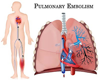
Brief Summary of Acute Pulmonary Embolism


Definition:

A pulmonary embolism results from the migration of venous thrombi from the systemic veins to pulmonary arterial system, resulting in varying degrees of obstruction of pulmonary arterial blood flow.
The incidence of pulmonary emboli in the United States exceeds 500,000 per year, with a mortality approaching 10%. If not diagnosed or if improperly treated, the mortality rate can reach 30%.

Common Sources:

Up to 90% of pulmonary emboli originate from the deep venous system of the legs. The upper extremities can also be a source of venous thrombi. Usually related to trauma, congenital fibromuscular bands, or the use of central venous catheters, 12% of all upper extremity thrombi result in pulmonary emboli. In addition, blood clot formation in the pelvic veins may cause either septic or bland pulmonary emboli, especially in the setting of complicated obstetric procedures or gynecologic surgery.
Other causes of pulmonary arterial obstructive emboli include air introduced during intravenous injections, hemodialysis, or the placement of central venous catheters; amniotic fluid secondary to vigorous uterine contractions; fat as a result of multiple long bone fractures; parasites; tumor cells; or injected foreign material (talc, mercury).

Risk Factors
:
Three basic risk factors, known collectively as Virchow’s triad, are associated with thrombus formation and subsequent pulmonary emboli:
  1. stasis,
  2. hypercoagulability, and 
  3. endothelial injury.

Management Of Acute Pulmonary Embolism


The goal of therapy is to prevent further embolic episodes, and heparin is the initial drug of choice for accomplishing this. First, a large intravenous loading bolus should be given, followed by continuous-drip infusion, maintained for at least 5 and often 7 to 10 days. Anticoagulation should not be withheld pending the results of further studies unless the patient’s risk of bleeding complications is greater than the clinical suspicion of pulmonary emboli. The partial thromboplastin time should be monitored and the heparin dosage adjusted to keep the time between 1.5 to 2.0 times the control.

Warfarin is started 24 to 48 hours after heparin therapy has been initiated. During the first 3 days of warfarin therapy, the prothrombin time or INR is increased before the onset of true anticoagulation. Therefore, before discontinuing the heparin, the prothrombin time or INR should be therapeutic (1.5 to 2 times normal) for approximately 2 to 3 days.

Low-molecular-weight heparins are indicated for prophylaxis in postoperative patients and probably have a role in the management of acute pulmonary embolism and deep venous thrombosis because they do not require monitoring of the anticoagulation effects.

Continuing the Treatment:
Long-term anticoagulation is usually achieved with warfarin, although low-molecular-weight heparins can also be used. Patients with reversible risk factors that are subsequently eliminated should undergo anticoagulation for a total of 3 months.

Monday, May 22, 2017

Introduction to Appendicitis


The most common abdominal surgical disease, appendicitis is inflammation of the vermiform appendix due to an obstruction. Although appendicitis may occur at any age and affects both sexes equally, it’s most common between puberty and age 30. Since the advent of antibiotics, the incidence and the death rate of appendicitis have declined. If left untreated, gangrene and perforation develop within 36 hours.

Causes
Appendicitis probably results from an obstruction of the intestinal lumen caused by a fecal mass, stricture, barium ingestion, or viral infection. This obstruction sets off an inflammatory process that can lead to infection, thrombosis, necrosis, and perforation. If the appendix ruptures or perforates, the infected contents spill into the abdominal cavity, causing peritonitis, the most common and most perilous complication of appendicitis.

Signs and symptoms
Appendicitis usually begins with generalized or localized colicky periumbilical or epigastric pain, followed by anorexia, nausea, and a few episodes of vomiting. Pain eventually localizes in the right lower quadrant of the abdomen (McBurney’s point), with abdominal “boardlike” rigidity, retractive respirations, increasing tenderness, increasingly severe abdominal spasms and, almost invariably, rebound tenderness. (Rebound tenderness on the opposite side of the abdomen suggests peritoneal inflammation.)
Later signs and symptoms include constipation (although diarrhea is also possible), slight fever, and tachycardia. Sudden cessation of abdominal pain indicates a perforation or infarction of the appendix. The elderly patient may present with minimal, vague symptoms and mild abdominal tenderness, delaying diagnosis of appendicitis.

Summary Points About Blepharitis


A common inflammation, blepharitis produces a red-rimmed appearance of the margins of the eyelids. In many cases, it’s chronic and bilateral and affects upper and lower lids. 

Seborrheic blepharitis is characterized by waxy scales and is common in older adults and in those with red hair. 

Staphylococcal (ulcerative) blepharitis is characterized by tiny ulcerated areas along the lid margins. 

Both types may coexist.
Blepharitis tends to recur and become chronic. It can be controlled if treatment begins before the onset of ocular involvement.
Causes
Seborrheic blepharitis generally results from seborrhea of the scalp, eyebrows, or ears; ulcerative blepharitis results from Staphylococcus aureus infection. (People with this infection may also tend to develop chalazions and styes.)
Signs and symptoms
Signs and symptoms of blepharitis include itching, burning, foreign-body sensation, and sticky, crusted eyelids on waking. This constant irritation results in unconscious rubbing of the eyes (causing reddened rims) or continual blinking. Other signs include waxy scales in seborrheic blepharitis; flaky scales on lashes, loss of lashes, and ulcerated areas on lid margins in ulcerative blepharitis.

Reye’s syndrome in Children


An acute childhood illness, Reye’s syndrome causes fatty infiltration of the liver with concurrent hyperammonemia, encephalopathy, and increased intracranial pressure (ICP). In addition, fatty infiltration of the kidneys, brain, and myocardium may occur.

Incidence
Reye’s syndrome affects children. It’s most common in patients ages 4 to 12, with a peak incidence at age 6.

Pathophysiology
Incidence of Reye’s syndrome usually rises during influenza outbreaks and is linked to aspirin use. It almost always follows within 1 to 3 days of an acute viral infection, such as an upper respiratory tract infection, type B influenza, or varicella (chickenpox).
With Reye’s syndrome, damaged hepatic mitochondria disrupt the urea cycle, which normally changes ammonia to urea for its excretion from the body. This results in hyperammonemia, hypoglycemia, and an increase in serum short-chain fatty acids, leading to encephalopathy.Simultaneously, fatty infiltration is found in renal tubular cells, neuronal tissue, and muscle tissue, including the heart.

Signs and symptoms
Reye’s syndrome develops in five stages, but the severity of the child’s signs and symptoms varies with the degree of encephalopathy and cerebral edema. Infants may have atypical presentation.
After the initial viral infection, a brief recovery period follows when the child doesn’t seem seriously ill. A few days later, he develops intractable vomiting, lethargy, rapidly changing mental status (mild to severe agitation, confusion, irritability, delirium), hyperactive reflexes, and rising blood pressure, respiratory rate, and pulse rate.
Reye’s syndrome may progress to coma. As the coma deepens, seizures develop, followed by decreased tendon reflexes and, commonly, respiratory failure.
Increased ICP, a serious complication, results from cerebral edema. Such edema may develop as a result of acidosis, increased cerebral metabolic rate, or an impaired autoregulatory mechanism.

Chronic Constipation - Causes , Signs&Symptoms And Management


Also known as lazy colon, colonic stasis, colonic inertia, and atonic constipation, chronic constipation may lead to fecal impaction if left untreated. It’s common in elderly and disabled people because of their inactivity and is commonly relieved with diet and exercise. Left untreated, it can result in hemorrhoids, fissures and megacolon.

Causes
Chronic constipation usually results from some deficiency in the three elements necessary for normal bowel activity: dietary bulk, fluid intake, and exercise. 
Other possible causes can include
  •  habitual disregard of the impulse to defecate, 
  • emotional conflicts, 
  • overuse of laxatives, or 
  • prolonged dependence on enemas, which dull rectal sensitivity to the presence of stool. 
Certain medications (tranquilizers, anticholinergics, opioids, antacids) can cause it, and patients with certain disorders (Parkinson’s disease, multiple sclerosis, hypothyroidism, scleroderma, lupus erythematosus) are more prone to develop it.

Signs and symptoms

The patient typically strains to produce dry, hard stool accompanied by mild abdominal discomfort. Straining can aggravate other rectal conditions such as hemorrhoids.

Brief Summary of Conjunctivitis


Hyperemia of the conjunctiva from infection, allergy, or chemical reactions characterizes conjunctivitis. Bacterial and viral conjunctivitis are highly contagious but are also self-limiting after 2 weeks. Chronic conjunctivitis may result in degenerative changes to the eyelids. In the Western hemisphere, conjunctivitis is probably the most common eye disorder.

Causes
The most common causative organisms are the following:
  • bacterial: Staphylococcus aureus, Streptococcus pneumoniae, Neisseria gonorrhoeae, Neisseria meningitidis
  • chlamydial: Chlamydia trachomatis (inclusion conjunctivitis)
  • viral: adenovirus types 3, 7, and 8; herpes simplex virus type 1.
Other causes include allergic reactions to pollen, grass, topical medications, air pollutants, and smoke; occupational irritants (acids and alkalies); rickettsial diseases (Rocky Mountain spotted fever); parasitic diseases caused by Phthirus pubis and Schistosoma haematobium; and, rarely, fungal infections.

Vernal conjunctivitis (also called seasonal or warm-weather conjunctivitis) results from allergy to an unidentified allergen. This form of conjunctivitis is bilateral; it usually begins before puberty and persists for about 10 years. Sometimes it’s associated with other signs and symptoms of allergy commonly related to grass or pollen sensitivity.
An idiopathic form of conjunctivitis may be associated with certain systemic diseases, such as erythema multiforme, chronic follicular conjunctivitis (orphan’s conjunctivitis), thyroid disease, and Stevens-Johnson syndrome. Conjunctivitis may be secondary to pneumococcal dacryocystitis or canaliculitis from candidal infection.

Sunday, May 21, 2017

Obsessive-compulsive disorder


Obsessive thoughts and compulsive behaviors represent recurring efforts to control overwhelming anxiety, guilt, or unacceptable impulses that persistently enter the consciousness.

The word obsession refers to a recurrent idea, thought, impulse, or image that is intrusive and inappropriate and causes marked anxiety or distress.

A compulsion is a ritualistic, repetitive, and involuntary defensive behavior. Performing a compulsive behavior reduces the patient’s anxiety and increases the probability that the behavior will recur. Compulsions are commonly associated with obsessions.

Patients with obsessive-compulsive disorder are prone to abuse psychoactive substances, such as alcohol and anxiolytics, in an attempt to relieve their anxiety. In addition, other anxiety disorders and major depression commonly coexist with obsessive-compulsive disorder.
Obsessive-compulsive disorder is typically a chronic condition with remissions and flare-ups. Mild forms of the disorder are relatively common in the population at large.

Causes

The cause of obsessive-compulsive disorder is unknown. Some studies suggest the possibility of brain lesions, but the most useful research and clinical studies base an explanation on psychological theories. Several studies show brain abnormalities, such as decreased caudal size and decreased white matter, but results are inconsistent and remain under investigation. In addition, major depression, organic brain syndrome, and schizophrenia may contribute to the onset of obsessive-compulsive disorder.

Introduction to Renal tubular acidosis


A syndrome of persistent dehydration, hyperchloremia, hypokalemia, metabolic acidosis, and nephrocalcinosis, renal tubular acidosis (RTA) results from the kidneys’ inability to conserve bicarbonate. This disorder occurs as distal RTA (type I, or classic RTA) or proximal RTA (type II). The prognosis is usually good but depends on the severity of renal damage that precedes treatment.

Causes
Metabolic acidosis usually results from renal excretion of bicarbonate. However, metabolic acidosis associated with RTA results from a defect in the kidneys’ normal tubular acidification of urine.

Distal RTA
Type I RTA results from an inability of the distal tubule to secrete hydrogen ions against established gradients across the tubular membrane. This results in decreased excretion of titratable acids and ammonium, increased loss of potassium and bicarbonate in the urine, and systemic acidosis.
Prolonged acidosis causes mobilization of calcium from bone and eventually hypercalciuria, predisposing the patient to the formation of renal calculi.
Distal RTA may be classified as primary or secondary:
Primary distal RTA may occur sporadically or through a hereditary defect and is most prevalent in females, older children, adolescents, and young adults.
Secondary distal RTA has been linked to many renal and systemic conditions, such as starvation, malnutrition, hepatic cirrhosis, and several genetically transmitted disorders.

Renal vein thrombosis


Clotting in the renal vein results in renal congestion, engorgement and, possibly, infarction. Renal vein thrombosis may affect both kidneys and may occur in an acute or a chronic form.
Chronic thrombosis usually impairs renal function, causing nephrotic syndrome. Abrupt onset of thrombosis that causes extensive damage may precipitate rapidly fatal renal infarction.
If thrombosis affects both kidneys, the prognosis is poor. However, less-severe thrombosis that affects only one kidney or gradual progression that allows development of collateral circulation may preserve partial renal function.

Causes
Renal vein thrombosis results from trauma to the abdomen or back, stricture (scar formation), or a tumor that obstructs the renal vein (usually hypernephroma).
Other causes include thrombophlebitis of the inferior vena cava (may result from abdominal trauma) or blood vessels of the legs, heart failure, and periarteritis. In infants, renal vein thrombosis usually follows diarrhea that causes severe dehydration.
Chronic renal vein thrombosis is a common complication of other glomerulopathic diseases, such as amyloidosis, systemic lupus erythematosus, diabetic nephropathy, and membranoproliferative glomerulonephritis.

Signs and symptoms
Signs and symptoms of renal vein thrombosis vary with the speed of onset.

Rapid onset of venous obstruction produces severe lumbar pain and tenderness in the epigastric region and the costovertebral angle. Other characteristic features include fever, leukocytosis, pallor, hematuria, proteinuria, peripheral edema and, when the obstruction is bilateral, oliguria and other uremic signs. The kidneys enlarge and become easily palpable. Hypertension is unusual but may develop.

Neonatal Resuscitation Procedure


Initial Steps

Prevent hypothermia—place infant on preheated radiant warmer, dry infant, then rewrap in warm, dry blankets. Polyethylene (food grade) bags may help maintain body temperature of very-low-birth weight infants.

Position—place the infant in a supine position with the head slightly extended (“sniffing position”); a small roll placed under the shoulders may be helpful.
Airway—if there are copious secretions compromising the airway, suction the mouth first and then the nose using either a suction bulb or 8F or 10F suction catheter; avoid prolonged and deep suctioning. Negative pressure should not exceed 100 mm Hg.
Tactile stimulation—if the infant remains apneic after drying, positioning, and suctioning, further tactile stimulation is unlikely to help; brief, gentle back rubbing or flicking the soles of feet, may be tried, but these efforts should not delay onset of positive-pressure ventilation.
Oxygen Administration
Free-flow 100% oxygen (at least 5 L/min) should be provided to any infant who has central cyanosis, pending further intervention(s). If positive-pressure ventilation (PPV) is begun, 100% supplemental oxygen is recommended. To reduce potential harm from excessive tissue oxygenation in preterm infants, the use of an oxygen blender and pulse oximetry is recommended in order to titrate supplemental oxygen delivery, maintaining oyxgen saturations between 90% and 95%. In any infant, if the heart rate does not respond by increasing to >100 beats per minute, 100% oxygen should be given.

Bag/Mask Ventilation

Indications—apnea or gasping; heart rate less than 100 beats per minute; central cyanosis despite free-flow oxygen.

Technique—proper mask size and shape, tight seal between mask and face, proper head position, clear airway, and adequate inflation pressure, ventilation rate of 40 to 60 breaths per minute. If ventilation pressure is being monitored, PPV with an initial inflation pressure of 20 cm H2O may be effective, but ?30 to 40 cm H2O may be required in some term babies without spontaneous ventilation.
Assessment—increasing heart rate is the primary sign of effective ventilation. Also look for improving color, spontaneous respirations, and improving muscle tone. If the heart rate is not improving, chest wall movement should be assessed and sufficient ventilating pressure should be used to move the chest wall with each breath.
Gastric decompression—if bag/mask ventilation is prolonged, an 8F orogastric tube should be inserted, aspirated, and left open as a vent to prevent gastric distension.
Special note: Flow-controlled pressure-limited mechanical devices (e.g., T-piece resuscitators) are recognized as an acceptable method of administering positive-pressure ventilation during newborn resuscitation; however, self-inflating and flow-inflating bags remain the preferred method for prolonged resuscitation.

Approach To A Patient Presenting With Haemoptysis (Blood In Sputum)


Haemoptysis (Blood In Sputum) may be the chief or sole complaint of patients presenting to the emergency department or their family doctors. . It always warrants investigation.

Causes of haemoptysis:

Respiratory causes;
• Infection (URTI, pneumonia, TB, lung abscess)
• Carcinoma (bronchial or laryngeal)
• Bronchiectasis

Cardiovascular causes:
• Pulmonary oedema
• PE
• Ruptured aortic aneurysm (aorto-bronchial fistula)

Coagulation disorder:
• Drugs (eg warfarin, heparin)
• Inherited (eg Haemophilia, Christmas disease)

Trauma:
• Penetrating or blunt

Others:
• Goodpasture’s, Wegener’s granulomatosis

Introduction to Osteoporosis


Introduction
In osteoporosis, a metabolic bone disorder, the rate of bone resorption accelerates while the rate of bone formation slows down, causing a loss of bone mass. Bones affected by this disease lose calcium and phosphate salts and thus become porous, brittle, and abnormally vulnerable to fracture.
Osteoporosis may be primary or secondary to an underlying disease. Primary osteoporosis is commonly called senile or postmenopausal osteoporosis because it’s most common in elderly, postmenopausal women.
Causes
The cause of primary osteoporosis is unknown; however, a mild but prolonged negative calcium balance, resulting from an inadequate dietary intake of calcium, may be an important contributing factor—as may declining gonadal adrenal function, faulty protein metabolism due to estrogen deficiency, and a sedentary lifestyle.
Causes of secondary osteoporosis include 
  • prolonged therapy with steroids or heparin, 
  • total immobilization or disuse of a bone (as with hemiplegia, for example), 
  • alcoholism, 
  • malnutrition,
  •  malabsorption,
  •  scurvy, 
  • lactose intolerance, 
  • hyperthyroidism, 
  • osteogenesis imperfecta, and 
  • Sudeck’s atrophy (localized to hands and feet, with recurring attacks).
Signs and symptoms
Osteoporosis is usually discovered when an elderly person bends to lift something, hears a snapping sound, and then feels a sudden pain in the lower back. Vertebral collapse, producing a backache with pain that radiates around the trunk, is the most common presenting feature. Any movement or jarring aggravates the backache.

Special considerations in Patients with Myocardial infarction


With myocardial infarction (MI), also known as heart attack, reduced blood flow through one of the coronary arteries results in myocardial ischemia and necrosis. With cardiovascular disease, the leading cause of death in the United States and western Europe, death usually results from the cardiac damage or complications of an MI.

Mortality is high when treatment is delayed; almost half of all sudden deaths due to an MI occur before hospitalization, within 1 hour of the onset of symptoms. The prognosis improves if vigorous treatment begins immediately.
  • Care for patients who have suffered an MI is directed toward detecting complications, preventing further myocardial damage, and promoting comfort, rest, and emotional well-being. Most MI patients receive treatment in the coronary care unit (CCU), where they’re under constant observation for complications.
  • On admission to the CCU, record the patient’s blood pressure, temperature, and heart and breath sounds, and monitor them regularly. Also, obtain an ECG.
  • Assess and record the severity and duration of pain; administer an analgesic. Avoid I.M. injections; absorption from the muscle is unpredictable.
  • Check the patient’s blood pressure after giving nitroglycerin, especially the first dose.
  • Frequently monitor the ECG to detect rate changes or arrhythmias. Place rhythm strips in the patient’s chart periodically for evaluation.
  • During episodes of chest pain, obtain ECG, blood pressure, and pulmonary artery catheter measurements for changes.
  • Watch for signs and symptoms of fluid retention (crackles, cough, tachypnea, and edema), which may indicate impending heart failure. Carefully monitor daily weight, intake and output, respirations, serum enzyme levels, and blood pressure.
  • Auscultate for adventitious breath sounds periodically (patients on bed rest frequently have atelectatic crackles, which may disappear after coughing) and for third or fourth heart sounds.
  • Organize patient care and activities to maximize periods of uninterrupted rest.

Saturday, May 20, 2017

Introduction to Endometriosis


Endometriosis  is defined as the presence of endometrial tissue outside the lining of the uterine cavity. Such ectopic tissue is generally confined to the pelvic area, most commonly around the ovaries, uterovesical peritoneum, uterosacral ligaments, and cul-de-sac, but it can appear anywhere in the body.
This ectopic endometrial tissue responds to normal stimulation in the same way the endometrium does. During menstruation, the ectopic tissue bleeds, causing the surrounding tissues to become inflamed. This inflammation causes fibrosis, leading to adhesions that produce pain and cause infertility.
Active endometriosis usually occurs between ages 30 and 40, but may be seen before age 20. Severe symptoms of endometriosis may have an abrupt onset or develop over many years. This disorder usually becomes progressively severe during the menstrual years; after menopause, it tends to subside.

Causes
The mechanisms by which endometriosis causes symptoms, including infertility, are unknown. The main theories to explain this disorder are:
  • transtubal regurgitation of endometrial cells and implantation at ectopic sites
  • coelomic metaplasia (repeated inflammation may induce metaplasia of mesothelial cells to the endometrial epithelium)
  • lymphatic or hematogenous spread to account for extraperitoneal disease.
Signs and symptoms
The classic symptom of endometriosis is acquired dysmenorrhea, which may produce constant pain in the lower abdomen as well as the vagina, posterior pelvis, and back. The pain usually begins 5 to 7 days before menses, reaches its peak on days of bleeding, and lasts for 2 to 3 days. It differs from primary dysmenorrheal pain, which is more cramplike and concentrated in the abdominal midline. The severity of pain doesn’t necessarily indicate the extent of the disease.

Tendinitis and Bursitis


A painful inflammation of tendons and of tendon-muscle attachments to bone, tendinitis usually occurs in the shoulder rotator cuff, hip, Achilles tendon, or hamstring.
Bursitis is a painful inflammation of one or more of the bursae—closed sacs that are lubricated with small amounts of synovial fluid that facilitate the motion of muscles and tendons over bony prominences. Bursitis usually occurs in the subdeltoid, olecranon, trochanteric, calcaneal, or prepatellar bursae.

Causes
Tendinitis commonly results from trauma (such as strain during sports activity), another musculoskeletal disorder (rheumatic diseases, congenital defects), postural misalignment, abnormal body development, or hypermobility.

Bursitis usually occurs in middle age from recurring trauma that stresses or pressures a joint or from an inflammatory joint disease (rheumatoid arthritis, gout). Chronic bursitis follows attacks of acute bursitis or repeated trauma and infection. Septic bursitis may result from wound infection or from bacterial invasion of skin over the bursa.

Signs and symptoms
Tendinitis and bursitis have characteristic signs and symptoms.

Introduction to Neurogenic bladder


Introduction
Also known as neuromuscular dysfunction of the lower urinary tract, neurologic bladder dysfunction, and neuropathic bladder, neurogenic bladder refers to all types of bladder dysfunction caused by an interruption of normal bladder innervation. Subsequent complications include urinary incontinence, residual urine retention, urinary tract infection, calculi formation, and renal failure. A neurogenic bladder can be spastic (hypertonic, reflex, or automatic), flaccid (hypotonic, atonic, nonreflex, or autonomous), or uncoordinated (dyssynergic).

Causes
At one time, neurogenic bladder was thought to result primarily from spinal cord injury; now, it appears to stem from a host of underlying conditions:
  • cerebral disorders, such as cerebrovascular accident, brain tumor (meningioma and glioma), Parkinson’s disease, multiple sclerosis, and dementia
  • spinal cord disease or trauma, such as spinal stenosis (causing cord compression) or arachnoiditis (causing adhesions between the membranes covering the cord), cervical spondylosis, myelopathies from hereditary or nutritional deficiencies and, rarely, tabes dorsalis
  • disorders of peripheral innervation, including autonomic neuropathies resulting from endocrine disturbances such as diabetes mellitus (most common)
  • metabolic disturbances, such as hypothyroidism, porphyria, or uremia (infrequent)
  • acute infectious diseases such as Guillain-Barré syndrome
  • heavy metal toxicity
  • chronic alcoholism
  • collagen diseases such as systemic lupus erythematosus
  • vascular diseases such as atherosclerosis
  • distant effects of cancer such as primary oat cell carcinoma of the lung
  • herpes zoster
  • sacral agenesis.
An upper motor neuron lesion (above S2 to S4) causes spastic neurogenic bladder, with spontaneous contractions of the detrusor muscles, elevated intravesical voiding pressure, bladder wall hypertrophy with trabeculation, and urinary sphincter spasms.

A lower motor neuron lesion (below S2 to S4) causes flaccid neurogenic bladder, with decreased intravesical pressure, increased bladder capacity and large residual urine retention, and poor detrusor contraction.

Thursday, May 18, 2017

Summary of Idiopathic Thrombocytopenic Purpura


Idiopathic thrombocytopenic purpura (ITP)
, thrombocytopenia that results from immunologic platelet destruction, may be acute (postviral thrombocytopenia) or chronic (Werlhof’s disease, purpura hemorrhagica, essential thrombocytopenia, autoimmune thrombocytopenia). Acute ITP usually affects children between ages 2 and 6; chronic ITP mainly affects adults younger than age 50, especially women between ages 20 and 40.

Causes
ITP may be an autoimmune disorder because antibodies that reduce the life span of platelets have been found in nearly all patients. The spleen probably helps to remove platelets modified by the antibody. Acute ITP usually follows a viral infection, such as rubella and chickenpox, and can follow immunization with a live virus vaccine. Chronic ITP seldom follows infection and is commonly linked to immunologic disorders, such as systemic lupus erythematosus or human immunodeficiency virus infection. It’s also linked to drug reactions.

Clinical Features
Signs and symptoms of ITP common to all forms of thrombocytopenia include petechiae, ecchymoses, and mucosal bleeding from the mouth, nose, and GI tract. Generally, hemorrhage is a rare physical finding. Purpuric lesions may occur in vital organs, such as the lungs, kidneys, or brain, and may prove fatal.
With acute ITP, which is common in children, onset is usually sudden and without warning, causing easy bruising, epistaxis, and bleeding gums. Onset of chronic ITP is insidious.

Introduction to Metabolic Acidosis


A physiologic state of excess acid accumulation and deficient base bicarbonate, metabolic acidosis is produced by an underlying pathologic disorder. Symptoms result from the body’s attempts to correct the acidotic condition through compensatory mechanisms in the lungs, kidneys, and cells.
Metabolic acidosis is more prevalent among children, who are vulnerable to acid-base imbalance because their metabolic rates are faster and their ratios of water to total body weight are lower. Severe or untreated metabolic acidosis can be fatal.

Etiology
Metabolic acidosis usually results from excessive burning of fats in the absence of usable carbohydrates. This can be caused by diabetic ketoacidosis, chronic alcoholism, malnutrition, or a low-carbohydrate, high-fat diet—all of which produce more keto acids than the metabolic process can handle.

Other causes include:
  • anaerobic carbohydrate metabolism: a decrease in tissue oxygenation or perfusion, as occurs with pump failure after myocardial infarction, or when pulmonary or hepatic disease, shock, or anemia forces a shift from aerobic to anaerobic metabolism, causing a corresponding rise in the lactic acid level
  • renal insufficiency and failure (renal acidosis): underexcretion of metabolized acids or the inability to conserve base
  • diarrhea and intestinal malabsorption: loss of sodium bicarbonate from the intestines, causing the bicarbonate buffer system to shift to the acidic side. For example, ureteroenterostomy and Crohn’s disease can also induce metabolic acidosis.
Less frequently, metabolic acidosis results from salicylate intoxication (overuse of aspirin), exogenous poisoning, or Addison’s disease with an increased excretion of sodium and chloride and the retention of potassium ions (due to a deficiency of glucocorticoids and mineralocorticoids).