Glaucoma occurs in several forms:
- chronic open-angle (primary),
- acute angle-closure,
- low tension (normal IOP that’s too high for a particular person),
- congenital (inherited as an autosomal recessive trait), and
- secondary to other causes.
Glaucoma is the second most common cause of blindness in the United States. About 2.5 million Americans are afflicted with the disease, but only 1 million know that they have it. Its incidence is highest among blacks, and it’s the single most common cause of blindness in that group. The visual prognosis is good with early treatment.
Causes
The cause of glaucoma varies according to the type of disorder:
Causes
The cause of glaucoma varies according to the type of disorder:
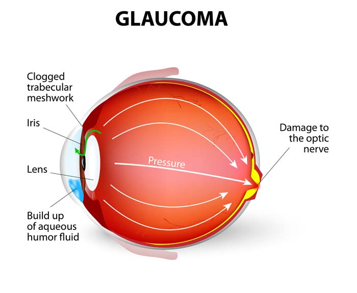
Chronic open-angle glaucoma results from overproduction of aqueous humor or from obstructed outflow of aqueous humor through the trabecular meshwork or the canal of Schlemm. This form of glaucoma frequently runs in families and affects 90% of all patients with glaucoma.
Acute angle-closure (narrow-angle) glaucoma results from obstructed outflow of aqueous humor caused by anatomically narrow angles between the anterior iris and the posterior corneal surface, shallow anterior chambers, a thickened iris that causes angle closure on pupil dilation, or a bulging iris that presses on the trabeculae, closing the angle. Adhesions in the angle, referred to as peripheral anterior synechiae, may be the cause.
Secondary glaucoma can result from uveitis, trauma, or drugs such as steroids. Neovascularization in the angle can result from vein occlusion or diabetes.
Signs and symptoms
Clinical features vary with the form of glaucoma.
Chronic open-angle glaucoma
Usually bilateral, chronic open-angle glaucoma has an insidious onset and a slowly progressive course. Symptoms appear late in the disease and include mild aching in the eyes, loss of peripheral vision, seeing halos around lights, and reduced visual acuity (especially at night) that’s uncorrectable with glasses.
Acute angle-closure glaucoma
An ophthalmic emergency, acute angle-closure glaucoma typically has a rapid onset. Symptoms include unilateral inflammation and pain, pressure over the eye, moderate pupil dilation that’s nonreactive to light, a cloudy cornea, blurring and decreased visual acuity, photophobia, and seeing halos around lights.
Because increased IOP may induce nausea and vomiting, glaucoma may be misinterpreted as GI distress. Unless treated promptly, this acute form of glaucoma produces blindness in 3 to 5 days.
Diagnosis
Loss of peripheral visual field, cupping of the optical disk, and increased IOP are the triad of signs that indicate glaucoma. Relevant diagnostic tests include the following:
- Tonometry (using an applanation, Schiøtz, or air-puff tonometer) measures IOP and provides a baseline for reference.
- Normal IOP ranges between 8 and 21 mm Hg, but some patients who fall in the normal range develop signs and symptoms of glaucoma. On the other hand, some patients who have abnormally high pressure have no clinical effects.
- Fingertip tension is another way to measure IOP. On gentle palpation of closed eyelids, one eye feels harder than the other in acute angle-closure glaucoma.
- Slit-lamp examination provides a look at the anterior structures of the eye, including the cornea, iris, and lens.
- Gonioscopy, by determining the angle of the anterior chamber of the eye, allows differentiation between chronic open-angle glaucoma and acute angle-closure glaucoma. The angle is normal in chronic open-angle glaucoma. In older patients, partial closure of the angle may also occur, so two forms of glaucoma may coexist.
- Ophthalmoscopy provides a look at the fundus, where cupping of the optic disk is visible in chronic open-angle glaucoma. This change appears later in chronic angle-closure glaucoma if the disease isn’t brought under control. A pale disk appears in acute angle-closure glaucoma.
- Perimetry or visual field tests help evaluate the extent of chronic open-angle deterioration by determining peripheral vision loss.
- Fundus photography can monitor the disk for any changes.
Advanced Centre For Eyes is known to provide the best eye treatment as we have an amazing eye specialist called Dr. Dinesh Garg who is famous for his finest treatment .
ReplyDelete